
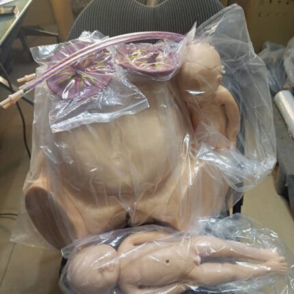
The Birthing Pelvis: Structure and Function in Childbirth
The female birthing pelvis serves as the crucial bony framework that enables successful vaginal delivery. This specialized structure combines anatomical precision with dynamic adaptability to facilitate the birth process.
Bony Architecture:
-
Composed of four interconnected bones:
-
Paired ilium bones (forming the sides)
-
Sacrum (posterior wall)
-
Coccyx (tailbone)
-
-
Forms a complete pelvic ring connecting the spine to the femurs
-
Provides structural support while allowing necessary movement
Morphological Features:
-
Distinctive hourglass configuration with:
-
Expanded false pelvis (greater pelvis) superiorly
-
Constricted true pelvis (lesser pelvis) inferiorly
-
Demarcated by the pelvic brim (superior strait)
-
-
Exhibits characteristic female adaptations:
-
Wider subpubic angle (>90°)
-
Broader, more circular inlet
-
Shorter sacral curvature
-
Obstetric Dimensions:
Three critical planes determine fetal passage:
-
Pelvic Inlet (Superior Strait):
-
Key diameter: Promontoretropubic (11.5-13 cm)
-
-
Midpelvis:
-
Crucial measurement: Median transverse (10.5 cm)
-
-
Pelvic Outlet:
-
Important span: Interspinous (10-11 cm)
-
Dynamic Adaptations:
-
Hormonally-mediated changes (relaxin, progesterone):
-
Increased sacroiliac joint mobility
-
Pubic symphysis widening (2-3 mm)
-
Ligamentous relaxation
-
-
Permits up to 10-15% expansion of birth canal dimensions
Parturition Mechanics:
-
Fetal Engagement: Head enters pelvic inlet
-
Descent: Progressive movement through planes
-
Rotation: Internal adjustment to navigate contours
-
Expulsion: Final passage through outlet


Reviews
There are no reviews yet.