
Adapted Tricycle
Adaptive Tricycle for Enhanced Mobility
DESCRIPTION:
This specially designed tricycle combines innovative engineering with user-friendly features to provide safe, comfortable mobility for individuals with limited movement.
Key Features:
✔ Modular Configuration
✔ Customizable Ergonomics
✔ Optimized Stability & Safety
✔ Dual Propulsion Options
✔ User-Centric Design

Air Compressor
RIXI-1EW Dental Air Compressor
*(Also known as ST-1EW model)*
Technical Specifications:
-
Power Supply: 220V AC / 50Hz | 2.4A
-
Output: 545W (545 VA)
-
Performance:
-
Flow Rate: 70 L/min
-
Working Pressure Range: 0.5–0.8 MPa
-
-
Construction: Heavy-duty metal housing for durability
-
Certification: CE compliant
Dental-Specific Features:
✔ Reliable Operation – Stable pressure output for dental handpieces and equipment
✔ Compact & Efficient – Optimized for clinical environments
✔ Low Maintenance – Robust metal construction ensures longevity

Anaerobic Jar
Materials:
Constructed primarily from durable stainless steel with a lid made of polycarbonate or PVC, both resistant to UV exposure and aging.
Capacity and Dimensions:
-
Volume: 2.5 liters
-
Holding Capacity: Accommodates up to 12 Petri dishes (90 mm in diameter)
Functionality:
Engineered to create and maintain anaerobic or microaerophilic conditions, enabling the cultivation of oxygen-sensitive microorganisms.
Pressure System:
-
Integrated valves for vacuuming air and introducing specific gases
-
Built-in pressure gauge for accurate control, typically ranging from -1 to +0.2 bar
Accessories and Compatibility:
-
Internal supports for Petri dishes, microtiter plates, or test tubes
-
Compatible with oxygen-removal systems such as palladium catalysts or gas-generating sachets
Application:
Essential in microbiology laboratories for the incubation and growth of anaerobic bacteria that require oxygen-free environments.

Anatomical Model of the Human Pelvis Skeleton
Anatomical Model of the Female Pelvis – Key Features
This detailed anatomical model accurately represents the structure and function of the female human pelvis, designed for both educational and clinical applications.
Bone Composition:
-
Includes paired iliac (coxal) bones, sacrum, and coccyx
-
Features L1 and L2 lumbar vertebrae to demonstrate spinal articulation
-
Provides complete representation of pelvic skeletal anatomy
Structural Characteristics:
-
Distinctive funnel-shaped architecture divided into:
-
Greater pelvis (false pelvis): Upper, flared portion
-
Lesser pelvis (true pelvis): Lower, narrower cavity
-
-
Anatomically accurate female morphology:
-
Wider, more circular pelvic inlet
-
Broader subpubic angle
-
Shorter, more everted sacrum
-
-
Designed to reflect obstetric adaptations for childbirth
Functional Attributes:
-
Supports weight transfer between trunk and lower limbs
-
Provides attachment points for major muscle groups
-
Protects reproductive organs and lower abdominal viscera
-
Facilitates bipedal locomotion and spinal stability
Educational & Clinical Applications:
-
Essential teaching tool for:
-
Medical and nursing education
-
Physical therapy programs
-
Midwifery training
-
-
Valuable clinical reference for:
-
Obstetric and gynecological practice
-
Orthopedic assessments
-
Surgical planning
-
-
Enables detailed study of:
-
Pelvic dimensions and variations
-
Biomechanical relationships
-
Pathological conditions
-

Anatomical Model of the Spine
The human spine anatomical model, commonly referred to as the spine, represents the central support structure of the human skeleton, extending from the skull’s base to the pelvis.
Structure and Composition:
Comprised of 33 vertically stacked vertebrae, separated by intervertebral discs that function as shock absorbers.
Segments:
Divided into five regions: 7 cervical vertebrae, 12 thoracic vertebrae, 5 lumbar vertebrae, the sacrum (5 fused vertebrae), and the coccyx (3-4 fused vertebrae).
Functions:
Serves three primary roles:
– **Static**: Maintains upright posture.
– **Dynamic**: Enables trunk and head mobility.
– **Protective**: Shields the spinal cord within the spinal canal.
Role:
Supports body weight, facilitates a wide range of movements, and acts as an anchor for muscles and ligaments.

Anatomy of the Human Urinary System
### **Human Urinary System – Key Features**
The urinary system consists of specialized organs that work together to filter blood, remove waste, and maintain fluid and electrolyte balance.
**Kidneys**
– Paired, bean-shaped organs that filter blood to eliminate waste (e.g., urea, excess salts).
– Regulate blood pressure, electrolyte levels, and red blood cell production.
**Ureters**
– Two narrow tubes that transport urine from the kidneys to the bladder.
**Bladder**
– A muscular, expandable sac that stores urine (capacity: ~600 mL).
– Contracts during urination to expel urine.
**Arteries & Veins**
– Represented in red (arteries) and blue (veins) on models.
– Supply blood to the kidneys for filtration and oxygenate urinary tissues.
**Urethra**
– The final passageway for urine excretion from the bladder to the exterior.

Autoclave
- Type: Portable, electrically heated steam autoclave for laboratory or medical use
- Material: High-quality SUS304 stainless steel
- Heating System: Electric heating system
- Leak-Proof: Silicone gasket for a secure, leak-proof seal
- Pressure Gauge: Dual-scale gauge to monitor working pressure
- Safety: Double safety valve for enhanced protection
- Tap: High-pressure tap included
- Volumes and Dimensions: Available in multiple volumes (e.g., 18L, 24L, 30L) with chamber sizes (e.g., Φ300-330mm for 18L, Φ300-460mm for 24L)
- Operating Temperature and Pressure: 115-129°C, 0.145-0.165 MPa
- Control: Features time (0-80 min) and temperature control, plus water shortage protection

Bedside Cabinet
Medical Bedside Cabinet – Hygienic Patient Storage Solution
Key Features:
✔ Clinical-Grade Construction
-
ABS plastic – Non-porous, chemical-resistant, and easy to disinfect
-
Smooth edges for patient safety
✔ Optimized Storage
-
2 drawers + 1 cabinet with door (organized medical/personal item storage)
-
Dimensions: 480×480×760mm (standard) or 450×410×740mm (compact)
✔ Mobility & Stability
-
4 swivel casters (2 with brakes) for easy repositioning
-
Lockable options available for secure medication storage

Bipolar Electrosurgical Unit
Bipolar Electrosurgical Unit – 400W Surgical Diathermy System
Key Features:
✔ High-Power Performance – Delivers 400W output for versatile surgical applications
✔ Multi-Mode Precision – 5 specialized modes:
-
Monopolar: Pure Cut, Blend Cut, Soft Coag, Forced Coag
-
Dedicated Bipolar Mode for controlled tissue effects
✔ Smart Safety Systems – -
Return electrode monitoring (burn prevention)
-
Automatic error detection & unipolar conversion
✔ Endoscopic Integration – -
Dedicated output port with auto-switching to endoscopic mode
Advanced Technology:
-
Digital Touchscreen – Real-time parameter visualization
-
Auto-Adjustment – Intelligent power adaptation for different tissues
Clinical Applications:
General | Thoracic | Orthopedic | Cardiac | Gynecological | Urological (incl. underwater TUR)
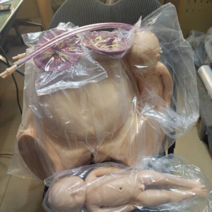
Birthing Pelvis with 2 Babies (Male and Female)
The birthing pelvis is the female bony structure essential for childbirth, providing both structural support and a dynamic passageway for fetal delivery.
Anatomical Composition
-
Comprises four fused bones:
-
Paired iliac bones (forming the pelvic girdle)
-
Sacrum (posterior base of the spine)
-
Coccyx (tailbone)
-
-
Connects the spine to the lower limbs, ensuring stability and weight distribution.
Shape & Structural Divisions
-
Funnel/hourglass shape with distinct regions:
-
Upper flare (false pelvis): Wider, supports abdominal organs.
-
Lower flare (true pelvis): Narrower, forms the birth canal.
-
-
Superior strait: The narrowing between these regions, a critical landmark for obstetric assessment.
Obstetric Straits & Key Measurements
The pelvis features three key straits, each with critical diameters for fetal passage:
-
Upper strait:
-
Promontoretropubic diameter (anteroposterior measurement).
-
-
Middle strait:
-
Median transverse diameter (widest point between ischial spines).
-
-
Lower strait:
-
Interspinous diameter (between ischial tuberosities).
-
-
These measurements determine cephalopelvic disproportion (CPD) and delivery feasibility.
Dynamic Adaptability
-
Not rigid: Hormonal changes (relaxin, progesterone) increase ligament laxity late in pregnancy.
-
Pubis symphysis and sacroiliac joints slightly widen, expanding the birth canal by up to 10–15%.
Role in Childbirth
-
Guides fetal descent: The baby navigates through the pelvic inlet, mid-cavity, and outlet.
-
Facilitates rotation: Bone contours help the fetus rotate into optimal positioning (e.g., occiput anterior).
-
Enables vaginal delivery: Optimal pelvic dimensions and adaptability prevent obstructed labor.

Birthing Pelvis with 2 Babies (Male and Female)
The Birthing Pelvis: Structure and Function in Childbirth
The female birthing pelvis serves as the crucial bony framework that enables successful vaginal delivery. This specialized structure combines anatomical precision with dynamic adaptability to facilitate the birth process.
Bony Architecture:
-
Composed of four interconnected bones:
-
Paired ilium bones (forming the sides)
-
Sacrum (posterior wall)
-
Coccyx (tailbone)
-
-
Forms a complete pelvic ring connecting the spine to the femurs
-
Provides structural support while allowing necessary movement
Morphological Features:
-
Distinctive hourglass configuration with:
-
Expanded false pelvis (greater pelvis) superiorly
-
Constricted true pelvis (lesser pelvis) inferiorly
-
Demarcated by the pelvic brim (superior strait)
-
-
Exhibits characteristic female adaptations:
-
Wider subpubic angle (>90°)
-
Broader, more circular inlet
-
Shorter sacral curvature
-
Obstetric Dimensions:
Three critical planes determine fetal passage:
-
Pelvic Inlet (Superior Strait):
-
Key diameter: Promontoretropubic (11.5-13 cm)
-
-
Midpelvis:
-
Crucial measurement: Median transverse (10.5 cm)
-
-
Pelvic Outlet:
-
Important span: Interspinous (10-11 cm)
-
Dynamic Adaptations:
-
Hormonally-mediated changes (relaxin, progesterone):
-
Increased sacroiliac joint mobility
-
Pubic symphysis widening (2-3 mm)
-
Ligamentous relaxation
-
-
Permits up to 10-15% expansion of birth canal dimensions
Parturition Mechanics:
-
Fetal Engagement: Head enters pelvic inlet
-
Descent: Progressive movement through planes
-
Rotation: Internal adjustment to navigate contours
-
Expulsion: Final passage through outlet

Burettes
Burettes – Precision Liquid Dispensing Instruments
(For Titrations & Volumetric Analysis)
1. Key Components
| Part | Description |
|---|---|
| Graduated Tube | Long cylindrical glass with precision markings (Class A/B tolerance) |
| Stopcock | PTFE or glass valve for flow control |
| Tip | Tapered delivery end for drop-wise dispensing |
| Mounting Clamp | For secure positioning on stands |
2. Technical Specifications
| Parameter | Details |
|---|---|
| Standard Volumes | 10 mL, 25 mL, 50 mL (Also available: 5 mL, 100 mL) |
| Graduation | 0.1 mL increments (50 mL) |
| Accuracy | ±0.05 mL (Class A) |
| Meniscus Reading | Bottom (clear liquids) / Top (dark solutions) |
| Material | Borosilicate glass (3.3) |
3. Types & Selection Guide
-
Digital Burettes: Motorized, programmable dispensing
-
Microburettes: For <1 mL volumes (0.01 mL gradations)
-
Automatic Zeroing: Self-refilling designs
4. Proper Usage Protocol
-
Rinsing: Pre-wet with titrant solution
-
Filling: Avoid air bubbles (use funnel)
-
Reading: Eye level at meniscus
-
Delivery: Slow, drop-wise near endpoint
5. Calibration & Maintenance
-
Verification: Annual calibration with distilled water
-
Cleaning: Chromic acid for stubborn residues
-
Storage: Vertical position, stopcock grease applied
