


Anatomical Model of the Spine
The human spine anatomical model, commonly referred to as the spine, represents the central support structure of the human skeleton, extending from the skull’s base to the pelvis.
Structure and Composition:
Comprised of 33 vertically stacked vertebrae, separated by intervertebral discs that function as shock absorbers.
Segments:
Divided into five regions: 7 cervical vertebrae, 12 thoracic vertebrae, 5 lumbar vertebrae, the sacrum (5 fused vertebrae), and the coccyx (3-4 fused vertebrae).
Functions:
Serves three primary roles:
– **Static**: Maintains upright posture.
– **Dynamic**: Enables trunk and head mobility.
– **Protective**: Shields the spinal cord within the spinal canal.
Role:
Supports body weight, facilitates a wide range of movements, and acts as an anchor for muscles and ligaments.

Anatomical Pelvis Model with 5 Lumbar Vertebrae
The main features of this anatomical pelvis model with 5 lumbar vertebrae are
Realistic and Life-Size Representation: This is a life-size model of the human pelvis, including the iliac bones, sacrum, coccyx, and the five lumbar vertebrae (L1 to L5), as well as the intervertebral discs.
Quality Materials: Made of high-quality PVC, it is durable and washable, and the pubic symphysis cartilage is made of rubber for increased realism.
Accurate Anatomical Details: The model preserves the details of each anatomical bone, including the left and right pelvic bones (ischium, ilium, and pubis) and the lumbar vertebrae with their structures.
Educational Functionality: Designed as a teaching and demonstration tool, it is ideal for the study of human anatomy, medical training, and communication between healthcare professionals and patients.
Mounting Stand: The model is mounted on a stable plastic base and includes a removable mounting stand for easy study and presentation.
Versatility: Suitable for use in a variety of settings such as education, art studies, cosmetics, and medical fields.

Anatomical Skin Model
Skin Characteristics
The skin, represented by the anatomical model, is the largest organ in the human body and has several essential characteristics and functions.
Structure
The skin is composed of three main layers, visible on the model:
The epidermis: The outermost layer, visible and palpable, which protects against toxins, bacteria, and fluid loss. It contains melanocytes that produce melanin, which filters UV rays.
The dermis: Located beneath the epidermis, it is a thick layer of elastic and fibrous tissue that gives the skin its strength and elasticity. It contains nerve endings, sweat and sebaceous glands, hair follicles, and blood vessels.
The hypodermis (or fatty layer): Located beneath the dermis, it insulates the body from heat and cold and serves as a protective layer for storing energy reserves. Functions
The skin performs many vital functions:
Protection: It protects the body from heat, cold, UV rays, injuries, and infections.
Thermal regulation: It controls body temperature through perspiration (produced by sweat glands) and vasodilation/vasoconstriction of blood vessels.
Sensory: It allows the perception of touch, pain, pressure, and temperature through nerve endings.
Vitamin D synthesis: Under the influence of sunlight, the skin participates in the production of vitamin D.
Excretion: It removes waste from the body through perspiration.
Aesthetic and psychosocial: Its color, texture, and quality play an important role in appearance and social interactions.

Anatomy of the Human Urinary System
### **Human Urinary System – Key Features**
The urinary system consists of specialized organs that work together to filter blood, remove waste, and maintain fluid and electrolyte balance.
**Kidneys**
– Paired, bean-shaped organs that filter blood to eliminate waste (e.g., urea, excess salts).
– Regulate blood pressure, electrolyte levels, and red blood cell production.
**Ureters**
– Two narrow tubes that transport urine from the kidneys to the bladder.
**Bladder**
– A muscular, expandable sac that stores urine (capacity: ~600 mL).
– Contracts during urination to expel urine.
**Arteries & Veins**
– Represented in red (arteries) and blue (veins) on models.
– Supply blood to the kidneys for filtration and oxygenate urinary tissues.
**Urethra**
– The final passageway for urine excretion from the bladder to the exterior.

Bandage Simulator
The features of the W44008 Surgical Bandage Simulator are as follows:
Simulator Type: This is a surgical bandage simulator, consisting of a female torso with natural elastic skin.
Main Function: It allows for realistic reproduction of adhesive bandage techniques and all forms of bandages.
Scars and Wounds: The simulator includes 14 different scars, as well as a mid-thigh stump after amputation (with surgical staples) for practicing treatment, cleaning, and bandaging techniques.
Accessories: It comes in a carrying case.
Technical Specifications: It weighs 11 kg and its dimensions are 84 x 30.5 x 63.5 cm.
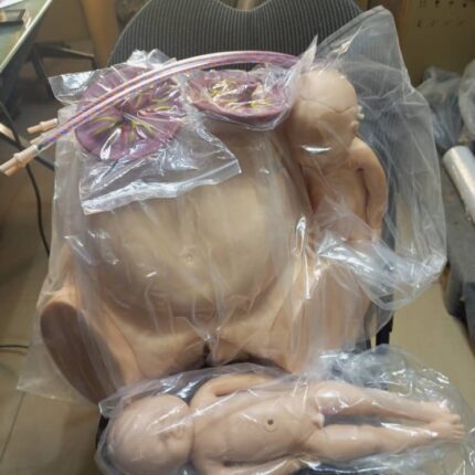
Birthing Pelvis with 2 Babies (Male and Female)
The birthing pelvis is the female bony structure essential for childbirth, providing both structural support and a dynamic passageway for fetal delivery.
Anatomical Composition
-
Comprises four fused bones:
-
Paired iliac bones (forming the pelvic girdle)
-
Sacrum (posterior base of the spine)
-
Coccyx (tailbone)
-
-
Connects the spine to the lower limbs, ensuring stability and weight distribution.
Shape & Structural Divisions
-
Funnel/hourglass shape with distinct regions:
-
Upper flare (false pelvis): Wider, supports abdominal organs.
-
Lower flare (true pelvis): Narrower, forms the birth canal.
-
-
Superior strait: The narrowing between these regions, a critical landmark for obstetric assessment.
Obstetric Straits & Key Measurements
The pelvis features three key straits, each with critical diameters for fetal passage:
-
Upper strait:
-
Promontoretropubic diameter (anteroposterior measurement).
-
-
Middle strait:
-
Median transverse diameter (widest point between ischial spines).
-
-
Lower strait:
-
Interspinous diameter (between ischial tuberosities).
-
-
These measurements determine cephalopelvic disproportion (CPD) and delivery feasibility.
Dynamic Adaptability
-
Not rigid: Hormonal changes (relaxin, progesterone) increase ligament laxity late in pregnancy.
-
Pubis symphysis and sacroiliac joints slightly widen, expanding the birth canal by up to 10–15%.
Role in Childbirth
-
Guides fetal descent: The baby navigates through the pelvic inlet, mid-cavity, and outlet.
-
Facilitates rotation: Bone contours help the fetus rotate into optimal positioning (e.g., occiput anterior).
-
Enables vaginal delivery: Optimal pelvic dimensions and adaptability prevent obstructed labor.

Birthing Pelvis with 2 Babies (Male and Female)
The Birthing Pelvis: Structure and Function in Childbirth
The female birthing pelvis serves as the crucial bony framework that enables successful vaginal delivery. This specialized structure combines anatomical precision with dynamic adaptability to facilitate the birth process.
Bony Architecture:
-
Composed of four interconnected bones:
-
Paired ilium bones (forming the sides)
-
Sacrum (posterior wall)
-
Coccyx (tailbone)
-
-
Forms a complete pelvic ring connecting the spine to the femurs
-
Provides structural support while allowing necessary movement
Morphological Features:
-
Distinctive hourglass configuration with:
-
Expanded false pelvis (greater pelvis) superiorly
-
Constricted true pelvis (lesser pelvis) inferiorly
-
Demarcated by the pelvic brim (superior strait)
-
-
Exhibits characteristic female adaptations:
-
Wider subpubic angle (>90°)
-
Broader, more circular inlet
-
Shorter sacral curvature
-
Obstetric Dimensions:
Three critical planes determine fetal passage:
-
Pelvic Inlet (Superior Strait):
-
Key diameter: Promontoretropubic (11.5-13 cm)
-
-
Midpelvis:
-
Crucial measurement: Median transverse (10.5 cm)
-
-
Pelvic Outlet:
-
Important span: Interspinous (10-11 cm)
-
Dynamic Adaptations:
-
Hormonally-mediated changes (relaxin, progesterone):
-
Increased sacroiliac joint mobility
-
Pubic symphysis widening (2-3 mm)
-
Ligamentous relaxation
-
-
Permits up to 10-15% expansion of birth canal dimensions
Parturition Mechanics:
-
Fetal Engagement: Head enters pelvic inlet
-
Descent: Progressive movement through planes
-
Rotation: Internal adjustment to navigate contours
-
Expulsion: Final passage through outlet

Complete Knee Solution
Zimmer Biomet’s NexGen Complete Knee Solution offers several notable features:
Implant Interchangeability: Femoral implants are interchangeable with pre-coated stemmed tibial components, A/P wedges, and trabecular metal tibial components.
Fixation Options: Cemented and cementless options are available for femoral, tibial, and patellar components.
High Range of Motion: CR Flex and LPS Flex components allow for a wide range of motion, with femoral flexion up to 125°-155°.
Materials: The implants are composed of non-ferromagnetic materials such as Zimaloy cobalt-chromium-molybdenum alloy, ultra-high molecular weight polyethylene, and titanium-aluminum-vanadium alloy (Ti-6Al-4V).
Kinematic Alignment: This system pioneered the concept of “kinematic alignment,” a surgical approach that aims to preserve the natural biomechanics of the joint by recreating the patient’s pre-arthritis knee motion patterns.
Modularity: The modular design of the NexGen Rotating Hinge (RH) allows for bone preservation and seamless intraoperative conversions from the NexGen Legacy Condylar Constrained Knee (LCCK).
Condylar Loading: The NexGen RH Knee’s femoral component and articulating surfaces are designed to maintain centralized contact throughout the entire range of motion, ensuring 95% condylar loading through the tibial condyles.

CPR Manikin
**Cardiopulmonary Resuscitation (CPR) Manikins**
CPR manikins are lifelike training tools designed to replicate human anatomy, enabling users to practice life-saving first aid techniques.
**Key Features:**
✔ **Realistic Anatomical Landmarks** – Includes clearly defined sternum and ribs for accurate hand placement during chest compressions.
✔ **Thoracic Simulation** – The rib cage offers human-like resistance to mimic real cardiac massage conditions.
✔ **Multiple Size Options** – Available in adult, child, and infant models to train for various emergency scenarios.
✔ **Quick Response (QCPR) Technology** – Advanced feedback system to monitor and improve CPR performance in real time.

Dental Anatomy Model
Features of the Dental Demonstration Model:
Materials:
Constructed from non-toxic, eco-friendly polyvinyl chloride (PVC) plastic.
Structure:
Features a full set of 32 fixed teeth, soft gums, and an included tongue for realistic representation.
Function:
Designed to demonstrate proper dental hygiene, especially correct toothbrushing techniques.
Use:
Serves as an effective educational tool for dental students, dentists (for patient instruction), and teachers (to promote good oral habits in school and kindergarten settings).
Simulation:
Boasts naturally colored gums and highly durable teeth with accurate anatomical shaping.
Durability & Maintenance:
Resistant to corrosion and easy to clean and disinfect for long-term use.

ECG Simulator
The ECG simulator offers several key features, ideal for training and testing medical equipment.
Simulation Versatility: It can simulate 12-lead ECG, adult and pediatric normal sinus rhythms, as well as other physiological parameters such as respiration, temperature, and multi-channel invasive pressure (IP)
Advanced Training Features: It provides training in precise electrode placement with anatomical landmarks and visual feedback on correct or incorrect placement.
Equipment Compatibility and Testing: Designed to test the proper functioning of ECG devices, monitors, recorders, and defibrillators, with the ability to administer defibrillation shocks via manikins or directly on the simulator.
Customization Capabilities: It displays ECG waveforms compliant with various standards and databases, or even user-defined waveform data.
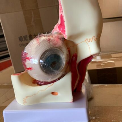
Eye and Orbit Anatomy
A magnified anatomical model of the human eye and orbit, designed for educational purposes in medical and academic settings.
Main Features:
- Magnification: Typically 3x the size of a real human eye and orbit, enhancing visibility of internal and external structures
- Composition and Detachability: Comprises multiple detachable parts (e.g., 10 parts), including the orbit, sclera, cornea, iris, ciliary body, choroid, and retina, allowing detailed study of the eye’s hierarchical structure
- Material: Made from high-quality, durable, and environmentally friendly PVC
- Fidelity and Detail: Features clear textures, high fidelity, and precise details to support accurate understanding of eye anatomy
- Educational Use: Ideal for teaching and studying eye anatomy, suitable for students and medical professionals