


Human Hand Anatomical Models
Human hand anatomical models possess several important features for the study of anatomy:
Detailed representation of structures: They illustrate the bones, muscles, ligaments, nerves, and arteries of the human hand, providing a comprehensive view of the anatomy.
Durable Materials: Made of durable PVC, these models are designed to be robust and durable, withstanding frequent use in educational settings.
muscles
Anatomical Accuracy: Developed by medical professionals, these models are designed to be 100% anatomically correct, making them ideal for teaching and learning.
Support Base: Most models are mounted on a stable base, facilitating observation and manipulation during study.
Educational Uses: They are valuable tools for medical training, classroom teaching, and improving patient communication, particularly when explaining conditions such as arthritis.

Anatomical Model of the Human Foot
The human foot is a complex anatomical structure with several important features, visible in models and confirmed by anatomy.
Key Features of the Human Foot:
Complex Bony Structure: The foot is composed of 26 bones, forming a longitudinal and transverse arch that acts as a shock absorber and support for the body’s weight.
Multiple Joints: It contains approximately 30 joints, allowing for great mobility and flexibility, particularly at the ankle, which connects the foot to the leg.
Muscles, Tendons, and Ligaments: More than 100 muscles, tendons, and ligaments work together to provide movement, stability, and support for the structure of the foot, including the arch.
Three Distinct Parts: The foot is subdivided into three main parts: the hindfoot (comprising the calcaneus and talus), the midfoot (comprising the cuboid, navicular, and cuneiform bones), and the forefoot (comprising the metatarsals and phalanges of the toes).
Essential Functional Role: The foot is fundamental for balance, locomotion (walking, running), and shock absorption during physical activities.
Form Variations: The models illustrate different foot shapes, such as the normal foot, flat foot (with a low arch), and pes cavus (with an excessively high arch), each with specific biomechanical implications.
Sensory Receptors: Sensory receptors detect touch, temperature, and pain, sending signals to the brain to adjust posture and gait.

Silicone IV Injection Practice Pad
The silicone IV injection practice pad has the following features:
Ultra-realistic material: Made of silicone, it mimics the texture and strength of human skin for a realistic tactile sensation during injections.
Simulated blood vessels: It typically incorporates four simulated blood vessels for practicing venipuncture and intravenous injections.
Durability and reusability: The skin and vessels can be punctured repeatedly without rapid deterioration, allowing for multiple training sessions.
Ease of filling: The vessels can be easily filled with water or blood-simulating liquid for a more realistic experience.
Training versatility: Suitable for practicing various techniques, including venipuncture, IV injection, infusion training, and blood transfusion.

Advanced Neonate Umbilical Cord Nursering Model
1. Female newborn systemic model , moveable head and limbs
2. Realistic umbilical cord with umbilical vein and artery
3. Umbilical cord ligation and nursering, realistic touch feeling ,the enough length of umbilical cord can be used continualy
4 . Neonatal basic nursery : lactation ,sponge Washing, wearing clothes, changing diapers

Complete Knee Solution
Zimmer Biomet’s NexGen Complete Knee Solution offers several notable features:
Implant Interchangeability: Femoral implants are interchangeable with pre-coated stemmed tibial components, A/P wedges, and trabecular metal tibial components.
Fixation Options: Cemented and cementless options are available for femoral, tibial, and patellar components.
High Range of Motion: CR Flex and LPS Flex components allow for a wide range of motion, with femoral flexion up to 125°-155°.
Materials: The implants are composed of non-ferromagnetic materials such as Zimaloy cobalt-chromium-molybdenum alloy, ultra-high molecular weight polyethylene, and titanium-aluminum-vanadium alloy (Ti-6Al-4V).
Kinematic Alignment: This system pioneered the concept of “kinematic alignment,” a surgical approach that aims to preserve the natural biomechanics of the joint by recreating the patient’s pre-arthritis knee motion patterns.
Modularity: The modular design of the NexGen Rotating Hinge (RH) allows for bone preservation and seamless intraoperative conversions from the NexGen Legacy Condylar Constrained Knee (LCCK).
Condylar Loading: The NexGen RH Knee’s femoral component and articulating surfaces are designed to maintain centralized contact throughout the entire range of motion, ensuring 95% condylar loading through the tibial condyles.
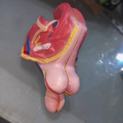
Male Genital Organ
This model consists of 4 parts and is life-size. It is easy to assemble and disassemble for dissecting the details of the internal and external structure of the male genital organ.
The 4 parts include:
2 halves of the penis showing median and transverse sections;
2 halves of the male genital apparatus.
With numbers to indicate:
1. Testicle
2. Epididymis
3. Ductus deferens; Vas deferens (sperm duct)
4. Seminal gland (Seminal vesicle)
5. Prostate
6. Bulbourethral gland (Cowper’s gland)
7. Penis
8. Scrotum
9. Corpora cavernosa of the penis
10. Spongy (cavernous) urethra (corpus cavernosum of the urethra)
11. Urinary bladder
12. Male urethra