


Kidney Anatomical Model
The Axis Scientific Human Kidney Anatomical Model features the following features:
Magnification:
It is magnified to 3 times life size, providing a detailed view of the kidney’s internal anatomy.
Detailed Anatomical Structures:
It displays the renal cortex, renal medulla (with renal pyramids), major and minor calyces, renal columns, renal pelvis, ureter, renal artery, and renal vein.
Adrenal Gland Included:
The model also includes the adrenal gland, located above the kidney.
Materials: It is generally made of high-quality, environmentally friendly, and durable PVC.
Structure Numbering:
The various anatomical parts are numbered and accompanied by a detailed manual to facilitate identification and learning.
Support Base:
The model is mounted on a stable, white base and can sometimes be removed for further study.
Educational Use:
It is designed as an ideal educational and teaching tool for anatomy and physiology students, as well as for healthcare professionals for patient education.

Anatomical Model of the Spine
The human spine anatomical model, commonly referred to as the spine, represents the central support structure of the human skeleton, extending from the skull’s base to the pelvis.
Structure and Composition:
Comprised of 33 vertically stacked vertebrae, separated by intervertebral discs that function as shock absorbers.
Segments:
Divided into five regions: 7 cervical vertebrae, 12 thoracic vertebrae, 5 lumbar vertebrae, the sacrum (5 fused vertebrae), and the coccyx (3-4 fused vertebrae).
Functions:
Serves three primary roles:
– **Static**: Maintains upright posture.
– **Dynamic**: Enables trunk and head mobility.
– **Protective**: Shields the spinal cord within the spinal canal.
Role:
Supports body weight, facilitates a wide range of movements, and acts as an anchor for muscles and ligaments.

Male Reproductive System Anatomical Model
The male reproductive system anatomical model offers a midsagittal view of the male pelvis, showcasing the structure and arrangement of the reproductive organs, bladder, and rectum.
Key Features:
Detailed Visualization:
The midsagittal section provides a clear, in-depth perspective of the male pelvis’s internal anatomy, including reproductive organs, bladder, and rectum.
Structures Highlighted:
Depicts key components such as the testes, epididymis, vas deferens, seminal vesicles, prostate, and urethra, among others.
Material Quality:
Constructed from durable PVC, ensuring accurate representation of anatomical details.
Educational Purpose:
Serves as a valuable medical education tool, enhancing understanding of the male reproductive system’s anatomy and physiology.
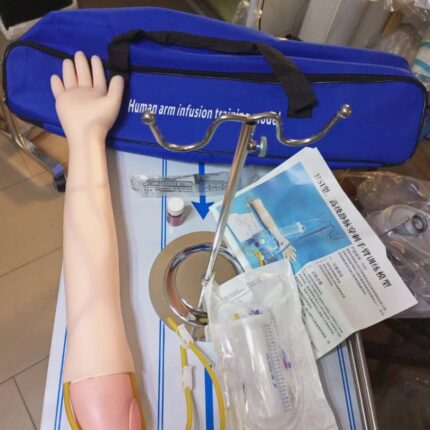
Injection Arm
The IV arm training model, like the one depicted, typically includes these features:
Realistic Design:
Mimics a human arm with adjustable skin and veins, offering a lifelike experience for practicing injections, blood draws, and infusions.
Authentic Puncture Feedback:
Replicates the sensation of needle insertion, including a distinct “pop” and simulated blood return upon correct vein puncture.
Durable and Reusable:
Constructed from high-quality materials, it endures hundreds of punctures without leaking, with self-sealing skin for repeated use.
Included Accessories:
Comes equipped with essential components for comprehensive training, such as an arm stand, fluid containers, and artificial blood.
Versatile Training:
Supports practice of multiple techniques, including IV injections, catheter placement, and blood collection.
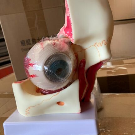
Eye and Orbit Anatomy
A magnified anatomical model of the human eye and orbit, designed for educational purposes in medical and academic settings.
Main Features:
- Magnification: Typically 3x the size of a real human eye and orbit, enhancing visibility of internal and external structures
- Composition and Detachability: Comprises multiple detachable parts (e.g., 10 parts), including the orbit, sclera, cornea, iris, ciliary body, choroid, and retina, allowing detailed study of the eye’s hierarchical structure
- Material: Made from high-quality, durable, and environmentally friendly PVC
- Fidelity and Detail: Features clear textures, high fidelity, and precise details to support accurate understanding of eye anatomy
- Educational Use: Ideal for teaching and studying eye anatomy, suitable for students and medical professionals
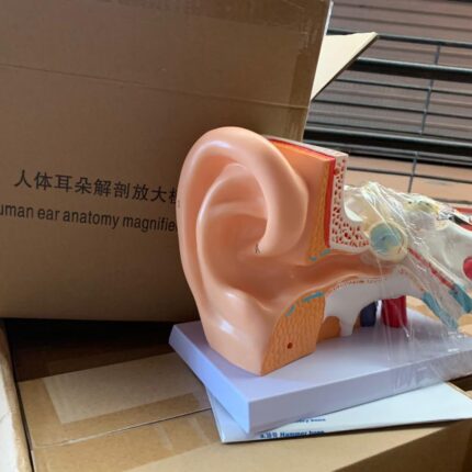
Human Ear Anatomical Model
A detailed, magnified model of the human ear designed for educational purposes in medical and academic settings.
Main Features:
- 5x Magnification: Enlarged five times the size of a real human ear for clear visualization of internal and external structures
- Detachable Design: Disassembles into at least two main parts (middle and inner ear) to facilitate study of overlapping elements
- Sturdy Material: Constructed from durable, wear-resistant PVC plastic with an ABS base for enhanced stability during display or teaching
- Labeled Structures: Precisely labeled parts, including outer ear, middle ear, inner ear, and cochlea, with color-coded sections for easy identification
- Versatile Use: Ideal for educational settings such as schools, hospitals, and laboratories, suitable for students, healthcare professionals, and educators