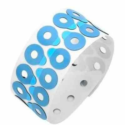
Adhesive tape
For Lens Processing:
Adhesion: Must securely bond with the blocking wax or alloy used for holding the lens.
Protection: Prevents scratches and damage to the lens surface during grinding and polishing.
Peelability: Must peel off cleanly and easily from the lens after processing, without leaving residue or transferring alloy.
Color: Often blue to make it easy to distinguish and remove from the lens.
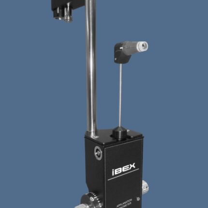
Applanation Tonometer
CT) use a puff of air to flatten the cornea, eliminating the need for anesthesia or contact.
Corneal Thickness Influence: The rigidity and thickness of the cornea affect the measurement; thinner corneas may result in artificially low readings, while thicker corneas may lead to falsely high ones.
Slit-Lamp Integration: The most common Goldmann applanation tonometer is mounted on a slit lamp.
Calibration: Instruments require daily calibration using standard weights to ensure accuracy.
Mire Rings (Goldmann): A split-image prism creates mires (marked rings) that are aligned to determine the applanation point and thus the IOP.
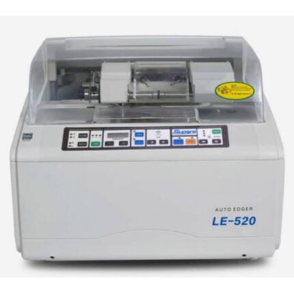
Auto Lens Edger
MODEL: LE-520
The LE-520 Auto Edger is an optical equipment designed for the automatic edger of eyeglass lenses.
Here are its main features:
Capable of processing CR39, glass, and PC (polycarbonate) lenses
Flat edge polishing and beveling functions
Equipped with a universal non-slip chuck
Standard PC functionality
Precision and Stability: Designed to process lenses with precision and stability
Ease of Use: Inherits the design style of Supore’s LE series, making it easy to use
Power Supply: 220V/50Hz
Unit Weight: Approx. 50 kg.
Power Consumption: 500W.
Technical Parameters (min/max):
Lens Size: Minimum 22 mm, Maximum 100 mm.
Adjustable Measuring Range: -6.0 mm to +6.0 mm in 0.05 mm increments.
Dimensions: 510 mm x 490 mm x 40 mm.
Grinding Wheels (Typical Configuration): Includes wheels for glass, CR, and polycarbonate, as well as a polishing wheel and a V-groove wheel.
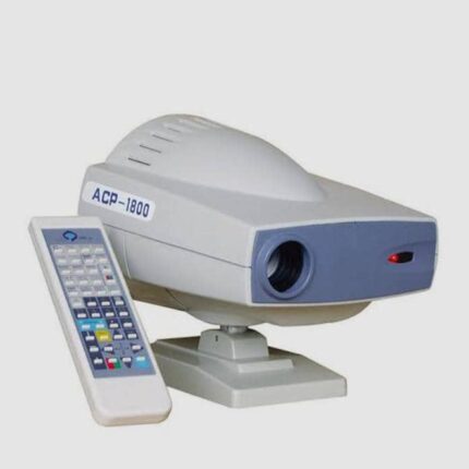
Automatic Chart Projector
MODEL: ACP-1800
The ACP-1800 Automatic Chart Projector is an ophthalmic instrument used for vision testing. Its main features are:
Projection Distance: It can project images at distances ranging from 1.5 m to 6 m.
Projection Magnification: The magnification is 30x at a distance of 5 m.
Projection Size: The projected image size is 330 mm (width) x 270 mm (height) at 5 m.
Number of Charts: It has 30 types of charts or graphs for vision testing.
Chart/Mask Change: Card or mask change is fast, taking 0.03 seconds per chart or mask image.
Lamps: It uses a 12V, 50W halogen lamp.
Programmability: Icons can be displayed in a predefined sequence via programming, and a single icon can be displayed via a multi-function mask plate.
Easy Installation and Maintenance: Bulb replacement is simple and requires no adjustment of focus or position.
Automatic Shutdown: The device automatically turns off after 10 minutes of inactivity.
Accessories: It comes with a remote control, a polarized metal shield, polarized glasses, fuses, and batteries.
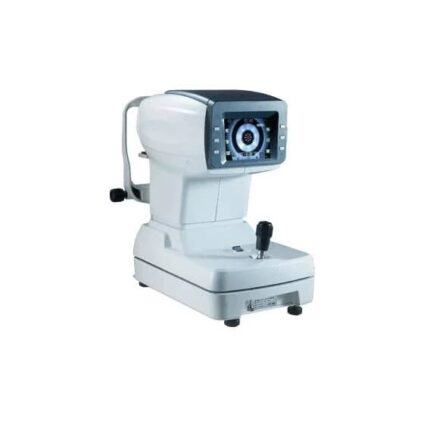
Autorefractometer
An autorefractometer is an ophthalmic device used to measure the optical characteristics of the eye, including refractive errors such as myopia, hyperopia, and astigmatism.
Key Features:
Refraction Measurement: It determines the sphere, cylinder, and axis of astigmatism for each eye.
Automation: Many models are fully automated, with features such as automatic alignment and measurement triggering.
Measurement Technology: Typically uses infrared light projected onto the retina to minimize accommodation and analyzes the reflected light to determine the necessary corrections.
Display and Interface: Equipped with a color LCD display (often tilting and rotating) and interfaces for connection to other equipment.
Keratometry: Some models, called autorefractokeratometers, also measure corneal radii of curvature and corneal astigmatism.
Speed and Accuracy: Allows rapid measurements (sometimes in seconds) with good accuracy, close to the final subjective refraction.
Additional Features: May include measurement of pupil diameter, interpupillary distance, and an optimized blurring system to minimize accommodation.
Children frame/trial frame
Lightweight and Comfortable:
Designed to be less burdensome and more comfortable on a child’s face, allowing for better cooperation during eye examination
Smaller Dimensions:
Smaller overall size and often fixed Pupillary Distance (PD) options, including specific infant and child sizes, to ensure a proper fit.
Adjustable Features:
Adjustable Temples: Allow for a customized length and fit to suit the child’s head size.
Lens Capacity:
Typically has multiple slots (often 3 or 4) to hold trial lenses and accessories like occluders or prisms.
Axis & PD Controls:
Knobs are present for rotating the cylindrical lens to adjust the astigmatism axis and for adjusting the interpupillary distance for accurate measurements.
Secure Fit Options:
An elastic headband can be included or used to help keep the frame centered on the child’s face.
Durable Materials:
Made from high-quality, durable, and long-lasting materials, often a combination of lightweight in
plastic and metal.
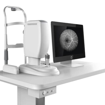
Color Fundus Camera
The device shown is a color fundus camera, a confocal ophthalmoscope for retinal angiography.
Its key features include:
Confocal laser scanning technology: Enables high-resolution imaging and stereoscopic viewing for depth perception.
High-resolution video capture: Ability to record videos at high resolutions, up to 1024 x 1024 pixels.
Multi-band fluorescence imaging and photography: Allows the use of different dyes such as fluorescein and indocyanine green to visualize retinal and choroidal vessels, although OCT angiography can reduce the need for dyes.
Non-mydriatic and continuous operation: Ability to capture images without prior pupil dilation, providing smoother operation.
Wide Field of View: Capable of capturing a 65-degree angle in a single undilated fundus photograph, with composite fields of view up to 120°.
High Resolution: Up to 1536 x 1536 pixels for high-resolution images.
Multiple Laser Sources: Uses different laser wavelengths (e.g., 488 nm for FA, 795 nm for ICGA, 830 nm for IR) for various imaging applications.
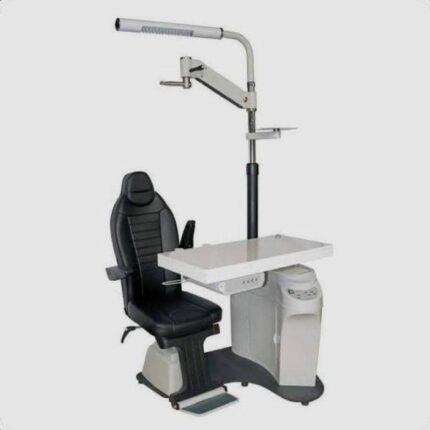
Complete Workstation Designed For Eye Examinations
The ophthalmic unit illustrated is a complete workstation designed for eye examinations, incorporating several key components:
Examination chair: A height-adjustable, often electrically powered, swiveling seat, providing comfort for the patient and flexibility for the ophthalmologist.
Instrument table: A lifting, sometimes sliding, table designed to accommodate various ophthalmic instruments such as a refraction head, an autorefractor, or a slit lamp.
Lighted column and supports: A vertical column incorporating an examination light (often halogen with adjustable lighting) and supports for other equipment, such as a chart projector or a motorized phoropter arm.
Control panel: Integrated controls on the table or a footswitch for adjusting the chair height and other unit functions.
Built-in storage: The unit often includes a cabinet or drawers for storing trial lenses and other small accessories.
Power supply: Built-in power transformers to power the instruments, as well as outlets for additional devices.
Ergonomic and compact design: Many units are designed to optimize space and facilitate intensive use in medical settings, with particular attention to stability and robustness.
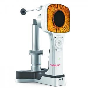
Digital portable slit lamp
Portability & Design: Lightweight and compact for convenient transport and use in mobile or remote settings.
Magnification: Offers 10x magnification with the option for 16x magnification.
Illumination: Equipped with a high-brightness white LED light source and a heat absorption filter.
Image Capture: Features a built-in camera and digital display for real-time, high-resolution image and video capture.
Display & Storage: Has a 3.5″ color screen and 8GB of internal storage for images.
User Interface: Manual focus mode and a user-friendly design for simple operation.
Connectivity: Includes Mini USB and WiFi for transferring data.
Power: Powered by a rechargeable 3.7V lithium battery, providing over 4 hours of continuous operation.
Slit Control: Allows for continuous adjustment of the slit width from 0 to 12mm.
Filters: Comes with red-free, heat absorption, and cobalt blue filters.
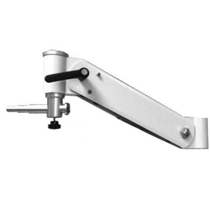
Electric chair/ Phoropter arm
Motorized Operation: Uses an electric mechanism for effortless and precise horizontal (and sometimes vertical) adjustments, reducing physical on the clinician.
Smooth and Stable Movement: Provides stable, controlled positioning of the phoropter, which is crucial for accurate ophthalmic measurements.
Durability: Constructed from high-quality, durable materials to ensure long-lasting performance in a clinical setting.
Precision Adjustment: Allows for fine-tuned, accurate positioning of the phoropter, improving the efficiency and precision of refractions.
Clinician Convenience: Designed for easy installation on compatible stands and examination units, improving workflow in a modern practice
Patient Comfort: Contributes to patient comfort by enabling smooth and effortless patient positioning.
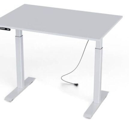
Electric Height-Adjustable Desk
- The image depicts an electric height-adjustable desk, also known as a sit-stand desk.
- It allows for easy switching between sitting and standing positions, helping to improve comfort and health at work.
- The height can be adjusted electrically, often with options to preset preferred heights.
- Using such a desk can help reduce muscle pain and joint problems, and promote better blood circulation.
- There are many models available, varying in size, materials, and features.
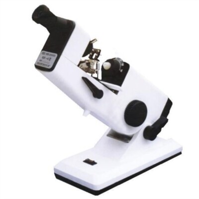
Hand lens meter
Lens power Measurement: Can measure spherical power, cylindrical power, and axis (direction) of cylindrical lenses.
Prism Power & Base Direction: Determines the strength of a prism and its base direction.
Optical Center Marking: Includes a precision inking attachment to mark the lens’s optical center for cutting and edging.
Used for quality control and manufacturing of lenses.
Opticians’ Shops: Essential for verifying prescription lenses and fitting glasses.
Ophthalmology: Employed in hospitals and clinics for comprehensive eye.
