
Intramedullary nails
The intramedullary nails shown, whether long or short, are orthopedic implants used to stabilize long bone fractures. Their main characteristics are:
Materials: They are generally made of titanium alloy or surgical stainless steel, biocompatible materials resistant to corrosion and mechanical stress.
Design:
Long nails: Designed for diaphyseal (central portion) fractures of long bones, such as the femur or tibia.
Short nails: Used for proximal fractures, such as those of the proximal humerus or proximal femur (subtrochanteric or intertrochanteric fractures).
Locking: They are equipped with locking screws (e.g., 4.5 mm) that pass through the bone and the nail to ensure stable fixation of the fracture and prevent rotation or shortening.
Functionality:
Cannulated: Many nails are cannulated, allowing for the insertion of a guide wire during surgery for precise positioning.
Indications: Their use varies depending on the patient’s age, the site, and the type of fracture, including diaphyseal and metaphyseal fractures, and nonunions.

Modular Revision Femoral Stem
The M-Vizion® Modular Revision Femoral Stem features the following:
Intelligent Modular Design: The M-Vizion system is designed with a modular approach to meet the challenges of femoral revision surgery, providing intraoperative flexibility.
Robust Modular Junction: The M-Vizion stem incorporates a proprietary mechanical design that improves the taper’s resistance to fretting-induced fractures, ensuring greater stability at the modular junction.
High mechanical strength of the taper and conical connection.
Diaphyseal Stability: The distal portion of the stem is based on the clinically proven Wagner concept, which provides immediate diaphyseal stability while avoiding areas of proximal bone loss.
Wagner-type cross-section with 8 longitudinal ribs for immediate stability and improved fixation.
Improved Proximal Fixation: The addition of a Mectagrip titanium plasma coating on the proximal body promotes proximal bone fixation.
Coatings and Surfaces:
TiNbN surface coating to prevent cold welding.
Grit Blasted Surface for long-term stability through bone fixation.
Specific Design Features:
Mirror-polished neck to minimize soft tissue damage and liner wear.
Rounded shoulder to facilitate greater trochanter reassembly.
Bullet-shaped tip for easier stem insertion.
Short stem with 12/14 taper to reduce the risk of liner wear.
3 holes for compatibility with cerclage cables and greater trochanter reconstruction.
Dimensions: Available in various lengths (e.g., 190 mm, 240 mm, 290 mm) and diameters (e.g., 12-26 mm), with proximal bodies of various sizes and offset options (standard and lateralized).
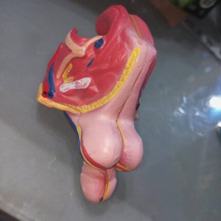
Male Genital Organ
This model consists of 4 parts and is life-size. It is easy to assemble and disassemble for dissecting the details of the internal and external structure of the male genital organ.
The 4 parts include:
2 halves of the penis showing median and transverse sections;
2 halves of the male genital apparatus.
With numbers to indicate:
1. Testicle
2. Epididymis
3. Ductus deferens; Vas deferens (sperm duct)
4. Seminal gland (Seminal vesicle)
5. Prostate
6. Bulbourethral gland (Cowper’s gland)
7. Penis
8. Scrotum
9. Corpora cavernosa of the penis
10. Spongy (cavernous) urethra (corpus cavernosum of the urethra)
11. Urinary bladder
12. Male urethra

Complete Knee Solution
Zimmer Biomet’s NexGen Complete Knee Solution offers several notable features:
Implant Interchangeability: Femoral implants are interchangeable with pre-coated stemmed tibial components, A/P wedges, and trabecular metal tibial components.
Fixation Options: Cemented and cementless options are available for femoral, tibial, and patellar components.
High Range of Motion: CR Flex and LPS Flex components allow for a wide range of motion, with femoral flexion up to 125°-155°.
Materials: The implants are composed of non-ferromagnetic materials such as Zimaloy cobalt-chromium-molybdenum alloy, ultra-high molecular weight polyethylene, and titanium-aluminum-vanadium alloy (Ti-6Al-4V).
Kinematic Alignment: This system pioneered the concept of “kinematic alignment,” a surgical approach that aims to preserve the natural biomechanics of the joint by recreating the patient’s pre-arthritis knee motion patterns.
Modularity: The modular design of the NexGen Rotating Hinge (RH) allows for bone preservation and seamless intraoperative conversions from the NexGen Legacy Condylar Constrained Knee (LCCK).
Condylar Loading: The NexGen RH Knee’s femoral component and articulating surfaces are designed to maintain centralized contact throughout the entire range of motion, ensuring 95% condylar loading through the tibial condyles.

Advanced Neonate Umbilical Cord Nursering Model
1. Female newborn systemic model , moveable head and limbs
2. Realistic umbilical cord with umbilical vein and artery
3. Umbilical cord ligation and nursering, realistic touch feeling ,the enough length of umbilical cord can be used continualy
4 . Neonatal basic nursery : lactation ,sponge Washing, wearing clothes, changing diapers

Silicone IV Injection Practice Pad
The silicone IV injection practice pad has the following features:
Ultra-realistic material: Made of silicone, it mimics the texture and strength of human skin for a realistic tactile sensation during injections.
Simulated blood vessels: It typically incorporates four simulated blood vessels for practicing venipuncture and intravenous injections.
Durability and reusability: The skin and vessels can be punctured repeatedly without rapid deterioration, allowing for multiple training sessions.
Ease of filling: The vessels can be easily filled with water or blood-simulating liquid for a more realistic experience.
Training versatility: Suitable for practicing various techniques, including venipuncture, IV injection, infusion training, and blood transfusion.

Anatomical Model of the Human Foot
The human foot is a complex anatomical structure with several important features, visible in models and confirmed by anatomy.
Key Features of the Human Foot:
Complex Bony Structure: The foot is composed of 26 bones, forming a longitudinal and transverse arch that acts as a shock absorber and support for the body’s weight.
Multiple Joints: It contains approximately 30 joints, allowing for great mobility and flexibility, particularly at the ankle, which connects the foot to the leg.
Muscles, Tendons, and Ligaments: More than 100 muscles, tendons, and ligaments work together to provide movement, stability, and support for the structure of the foot, including the arch.
Three Distinct Parts: The foot is subdivided into three main parts: the hindfoot (comprising the calcaneus and talus), the midfoot (comprising the cuboid, navicular, and cuneiform bones), and the forefoot (comprising the metatarsals and phalanges of the toes).
Essential Functional Role: The foot is fundamental for balance, locomotion (walking, running), and shock absorption during physical activities.
Form Variations: The models illustrate different foot shapes, such as the normal foot, flat foot (with a low arch), and pes cavus (with an excessively high arch), each with specific biomechanical implications.
Sensory Receptors: Sensory receptors detect touch, temperature, and pain, sending signals to the brain to adjust posture and gait.

Human Hand Anatomical Models
Human hand anatomical models possess several important features for the study of anatomy:
Detailed representation of structures: They illustrate the bones, muscles, ligaments, nerves, and arteries of the human hand, providing a comprehensive view of the anatomy.
Durable Materials: Made of durable PVC, these models are designed to be robust and durable, withstanding frequent use in educational settings.
muscles
Anatomical Accuracy: Developed by medical professionals, these models are designed to be 100% anatomically correct, making them ideal for teaching and learning.
Support Base: Most models are mounted on a stable base, facilitating observation and manipulation during study.
Educational Uses: They are valuable tools for medical training, classroom teaching, and improving patient communication, particularly when explaining conditions such as arthritis.

Human Trachea Intubation Model
Features
1. Accurate, full-fledged anatomy of the mouth, tongue, airway, and esophagus. Perfect for moving endotracheal tubes.
2. Respiratory auscultation.
3. Oral and nasal intubation.
4. Oral and nasal suction.
5. Soft neck with crocodile cartilage allows for classic Sellick maneuvering, necessary to provide a better view of the vocal cord.

Anatomical Model of a Monocot Stem
Characteristics of a Monocot Stem
The characteristics of a monocot stem, as represented by the model and described in the sources, include the following:
Epidermis: The outermost layer, covered by a thick cuticle and capable of bearing trichomes and stomata.
Hypodermis: The layer beneath the epidermis, generally composed of sclerenchyma cells.Scattered throughout the ground tissue in several concentric circles, each bundle is surrounded by a sheath of sclerenchyma cells.
Closed Crybrovascular Bundles: Unlike dicots, the vascular bundles of monocots are closed, meaning they lack fascicular or interfascicular cambium and do not allow for secondary thickness growth.
Xylem and Phloem: In each bundle, the xylem is endoarch (inward) and the phloem is outward. Xylem consists of vessels and parenchyma, while phloem includes sieve tubes, companion cells, and parenchyma.
Lack of Distinct Pith: The center of the stem is occupied by parenchyma cells, without a distinct pith as in dicots.

ECG Simulator
The ECG simulator offers several key features, ideal for training and testing medical equipment.
Simulation Versatility: It can simulate 12-lead ECG, adult and pediatric normal sinus rhythms, as well as other physiological parameters such as respiration, temperature, and multi-channel invasive pressure (IP)
Advanced Training Features: It provides training in precise electrode placement with anatomical landmarks and visual feedback on correct or incorrect placement.
Equipment Compatibility and Testing: Designed to test the proper functioning of ECG devices, monitors, recorders, and defibrillators, with the ability to administer defibrillation shocks via manikins or directly on the simulator.
Customization Capabilities: It displays ECG waveforms compliant with various standards and databases, or even user-defined waveform data.

Advanced Trauma Nursing Manikin
The Advanced Trauma Nursing Manikin, as shown in the image, features various features designed for medical and nursing training, including trauma simulation and nursing practice.
Key Features:
Various Trauma Simulation: The manikin is equipped with modules simulating various injuries and traumatic conditions, such as facial burns (grades I, II, and III), forehead lacerations, jaw wounds, open fractures (clavicle, humerus, femur, tibia), gunshot wounds, abdominal eviscerations, and amputations.
Nursing Capabilities: It allows for practicing a wide range of nursing procedures, including washing, disinfection, hemostasis, bandaging, and securing wounds, as well as holistic care such as sponge bathing and changing clothes.
Advanced Medical Procedures: The manikin facilitates training for more complex procedures such as endotracheal intubation, tracheostomy, injections (subcutaneous, intramuscular, intravenous), thoracic, abdominal, hepatic, and lumbar puncture, as well as ostomy care and urinary catheterization.
Realistic Anatomy: Designed with accurate anatomical details, it offers a realistic learning experience for trauma assessment and airway management, including a normal and dilated eye.
Durable and Hygienic Materials: Made of high-quality PVC, it is corrosion-resistant, odorless, easy to clean, and durable, ensuring a long lifespan for intensive educational use.
Modularity and Interchangeability: Some parts, such as skin burns or genitals, can be replaced, allowing you to simulate different scenarios and adapt to training needs.
