
Ultrasound Biomicroscopy System
The characteristics of an ultrasound biomicroscopy (UBM) system like the one shown include several key aspects, including technology, applications, and benefits.
Technology and Specifications:
High Frequency: Uses very high-frequency ultrasound (e.g., 50 MHz) for high-resolution imaging of the anterior segment of the eye.
Resolution and Penetration: Provides very high-resolution cross-sectional images with good tissue penetration, allowing visualization of fine structures such as the cornea, iris, ciliary body, and iridocorneal angle.
Portability: Often designed to be portable, connecting to laptops or desktops via USB, facilitating its use in various clinical environments.
Integrated Software: Works with dedicated software for image acquisition, analysis, and management, often with measurement and reporting features.
Specialty Probes: Uses sheathless oscillating probes designed for ocular examination, sometimes with guards and alarms for patient safety.
Clinical Applications:
Anterior Segment Analysis: Allows for detailed examination of the cornea, lens, ciliary body, iris, and iridocorneal angle.
Glaucoma Diagnosis: Essential for analyzing iridocorneal angle opening and detecting angles at risk of closure.
Evaluation of Ocular Pathologies: Used in the diagnosis and follow-up of various conditions, including ocular trauma, ciliary body disease, and other anterior segment pathologies.
Postoperative Follow-up: Allows for screening for complications after filtration surgery and assessment of reasons for surgical failure.
Benefits:
Detailed Imaging: Provides clear visualization of ocular structures difficult to observe with other methods. Non-invasive: UBM is a non-invasive procedure for analyzing the anterior segment of the eye.
Versatility: Applicable in a wide range of clinical situations for diagnosis, monitoring, and research in ophthalmology.

Phacoemulsifier
MODEL: MD-480A
The Meda MD-480A Phacoemulsifier features:
Control and Monitoring: All parts of the system are fully controlled and monitored by microprocessor technology.
Ultrasonic Emulsification (U/S):
Phaco Emulsification Frequency: 40 kHz ± 20%.
U/S Handpiece Upper Tip Vibration Accuracy: Max. 120 μm ± 20%.
Operating Modes: Continuous, Pulse, Linear, Burst.
Emulsification Power: 0-100%.
Pulse Frequency: 1 Hz to 99 Hz.
Burst Time: 5 ms to 100 ms.
Irrigation/Aspiration (I/A):
Peristaltic pump for aspiration.
Maximum flow rate: 40 ml/min.
Reflux flow rate: 20 ml/min.
Maximum vacuum: 66.7 kPa (500 mmHg).
Vitrectomy:
Cutting frequency: 20 to 600 cuts/min.
Cutting power source: Air pump.
Cauterization:
Frequency: 1 MHz.
Maximum power: 10 watts (with a 200 Ω load).
Power range: 0-100%.
User interface: Large TFT touchscreen and user-friendly menu.
Additional features:
Up to 10 sets of preset parameters.
Automatic testing for tubing and handpiece problems.
Intelligent voice reminder.
Electrical Requirements: 220 Volts ± 10%, 50 Hz ± 2%.
Dimensions and Weight: 43 cm (W) × 22.5 cm (H) × 32 cm (D), Weight: 8 kg.

Matronix B-scan Biometer
The Matronix B-scan Biometer is an advanced ophthalmic ultrasound system designed for the diagnosis and monitoring of ocular conditions. Its key features include:
High-resolution imaging: Provides clear and detailed images of internal ocular structures, with 256-level grayscale imaging for exceptional tissue differentiation.
Probes: Typically equipped with a 10 MHz probe for accurate measurements.
Display: Incorporates a large 10.4-inch or 12.1-inch LED display for comfortable viewing and easy image interpretation.
Scanning modes: Combines A-scan and B-scan modes, allowing for distance measurements (ACD, lens, vitreous, AXL) and observation of intraocular and posterior segment diseases.
Portability and Design: Compact and portable tabletop model (dimensions: 256 x 150 x 326 mm; weight: 4.5 kg), ideal for clinics and mobile environments.
Power Supply: Mains powered (AC 100-240V) and often equipped with a 4400 mAh lithium battery for stand-alone use.
Additional Features: Supports single and multiple image selection, real-time dynamic scanning to display lesion location, volume, and shape, and can be connected to a video recorder or ultrasound workstation.
Clinical Applications: Used for the diagnosis of ocular and orbital diseases, including refractive media turbidity, ocular somatometry, pre- and post-vitrectomy diagnosis, intraocular foreign body localization, intraocular tumors, exophthalmos, and ocular and orbital hemodynamics.

Ophthalmic YAG Laser System
MODEL: MD-920
The MD-920 ophthalmic YAG laser system, as shown in the image, has the following specifications:
Laser Specifications:
Laser Type: Nd:YAG, Q-switched
Wavelength: 1064 nm
Mode: Q-switched (multimode)
Pulse Output: Single pulse, double pulse, and triple pulse burst
Pulse Duration: 4.5 ns
Repetition Rate: 2.5 Hz
Cone Angle: 18°
Spot Size: 30 µm
Aiming Beam Wavelength: 633 nm
Aiming Beam Output Power: <0.4 mW
Slit Lamp (Galilean Stereoscopic Microscope) Features:
Provides high clarity and sharpness, avoiding double images.
Five-level drum magnification.
Optional accessories: teaching tube, CCD camera, and tonometer.
Other Features:
Easy setup for installation and maintenance.
Mounted on a mobile rolling stand for transport.
Uses microprocessor control technology with a digital display for easy operation.
Built-in laser protection system to ensure surgical safety.
Color Fundus Camera
The device shown is a color fundus camera, a confocal ophthalmoscope for retinal angiography.
Its key features include:
Confocal laser scanning technology: Enables high-resolution imaging and stereoscopic viewing for depth perception.
High-resolution video capture: Ability to record videos at high resolutions, up to 1024 x 1024 pixels.
Multi-band fluorescence imaging and photography: Allows the use of different dyes such as fluorescein and indocyanine green to visualize retinal and choroidal vessels, although OCT angiography can reduce the need for dyes.
Non-mydriatic and continuous operation: Ability to capture images without prior pupil dilation, providing smoother operation.
Wide Field of View: Capable of capturing a 65-degree angle in a single undilated fundus photograph, with composite fields of view up to 120°.
High Resolution: Up to 1536 x 1536 pixels for high-resolution images.
Multiple Laser Sources: Uses different laser wavelengths (e.g., 488 nm for FA, 795 nm for ICGA, 830 nm for IR) for various imaging applications.

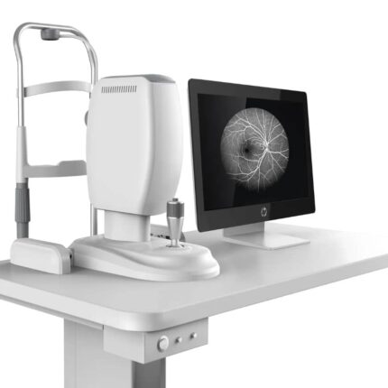
Optical Coherence Tomograph
The device shown is a Nidek RS-3000 Advance 2 optical coherence tomograph (OCT). Its key features include:
Comprehensive Retinal and Glaucoma Analysis: It offers a complete solution for analyzing retinal pathologies and glaucoma.
SLO-Based Eye Tracker: A scanning laser ophthalmoscope (SLO)-based eye tracker system enables precise image capture with eye movement compensation.
Selectable OCT Sensitivity: Enables B-scan image acquisition even through opacities in the ocular media.
HD Plus Tracing: This feature ensures accurate averaging of up to 120 images for high-resolution, high-contrast images.
OCT Angiography (OCT-A): High-resolution OCT angiographic imaging capability for observing microvascular details of the retina.
Glaucoma Analysis with Normative Database: Includes glaucoma analysis with an extensive normative database (9 x 9 mm).
High Scanning Speed: The device can reach 85,000 A-scans per second, enabling rapid image acquisition.
Automatic Panoramic Imaging: Automatic panoramic imaging capability for OCT angiography up to 12 x 12 mm.
Vibrant Color LCD Display: Equipped with an 8.4-inch color LCD display for real-time data viewing and review.
Comfortable Working Distance: A 35.5 mm working distance ensures comfortable patient positioning.

Ophthalmic Operating Table
The features of an ophthalmic operating table, such as the one shown, generally include the following:
Specialized Design for Ophthalmology: The table is specifically designed for ophthalmic surgeries and treatments, providing optimal positioning for delicate ocular procedures.
Triple-Section Tabletop: The tabletop is often divided into three sections, with a detachable head section and an integrated drainage system to facilitate eye surgeries.
Durable and Easy-to-Clean Materials: The base and upholstery are made of stainless steel, ensuring good stability, corrosion resistance, and easy cleaning, thus reducing the risk of infection.
Ergonomic Adjustments: The table allows for various position adjustments (height, Trendelenburg, lateral tilt) for patient comfort and surgeon ergonomics, often controlled by a manual unit.
Special Accessories: It is equipped with an ophthalmic headrest for stable positioning of the patient’s head and can include padded arm supports for the surgeon.
Operation: Available in hydraulic or electric options, providing smooth and precise tabletop movement for precise patient positioning.

Non-Contact Tonometer
The NCT-100 non-contact tonometer is an ophthalmic device designed for measuring intraocular pressure (IOP) using a puff of air.
Its main features are:
Fully Automatic Measurement: It allows for fully automatic measurement of data for both eyes without the need for manual switching.
Automatic 3D Positioning: Automatic 3D (xyz) positioning ensures accurate and reliable measurements.
Ease of Use: The device is easy to use thanks to its color touchscreen.
Data Stability: It provides stable IOP data thanks to a gentle puff of air measurement.
Corneal Thickness Compensation: It is capable of compensating for corneal thickness during IOP measurement.
Speed: It provides measurement results in less than 10 seconds.
Non-contact: The non-contact measurement method reduces the risk of eye injury or infection.
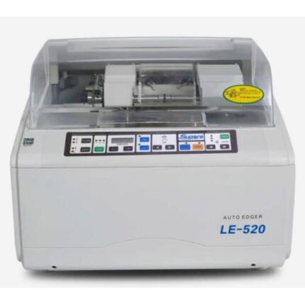
Auto Lens Edger
MODEL: LE-520
The LE-520 Auto Edger is an optical equipment designed for the automatic edger of eyeglass lenses.
Here are its main features:
Capable of processing CR39, glass, and PC (polycarbonate) lenses
Flat edge polishing and beveling functions
Equipped with a universal non-slip chuck
Standard PC functionality
Precision and Stability: Designed to process lenses with precision and stability
Ease of Use: Inherits the design style of Supore’s LE series, making it easy to use
Power Supply: 220V/50Hz
Unit Weight: Approx. 50 kg.
Power Consumption: 500W.
Technical Parameters (min/max):
Lens Size: Minimum 22 mm, Maximum 100 mm.
Adjustable Measuring Range: -6.0 mm to +6.0 mm in 0.05 mm increments.
Dimensions: 510 mm x 490 mm x 40 mm.
Grinding Wheels (Typical Configuration): Includes wheels for glass, CR, and polycarbonate, as well as a polishing wheel and a V-groove wheel.

Optical Lens Cutting Machine
MODEL: SJG-1000
The features of the SJG-1000 (or SJG-1000AK) optical lens cutting machine are as follows:
Tracing and Scanning System:
Optical scanning and centering are positioned as a single system.
Obtain the frame shape in 0.5 seconds.
Real-time imaging guidance to confirm the optimal center position.
Automatic elimination of lens, prism, and artificial parallax effects.
Display and Interface:
7-inch high-resolution (800*600) color touch screen.
Simple operation and low failure rate, requiring little maintenance.
Lens Processing Capabilities:
Suitable for various lens materials, including glass, CR, PC, high-index lenses, and CR-39.
Equipped with various wheels (diamond for glass, V-groove for CR/PC, bevel polishing).
Processes lenses from 22 mm to 80 mm.
Automatic and manual beveling are available.
Grooving width, depth, and position are manually adjustable.
V-shaped and flat polishing are available.
Other Features:
Can be connected simultaneously to two SJG-1000AK automatic milling machines to form a processing center.
3D plotter for accurate lens imaging in 1 second on an 8-inch monitor (for the AK version).
Built-in, easy-to-use blocker (for the AK version).
Four-level adjustable scanning light source (for the AK version).
Compact design for easy placement in a workshop or laboratory.

Lens Edger
Modèle :NAME: Lens Edger
The ALE-1000 Auto Lens Edger is a template-free lens edger with several notable features.
Key Features:
Screen and User Interface:
5.7-inch color TFT LCD screen with a high resolution of 640×480.
1:1 display of lens graphics, icons, and a user-friendly interface.
Design and Ergonomics:
Ergonomic design with a sleek shape.
Divided configuration between the main body and scanners.
Lens Processing Capabilities:
Supports various lens types, including CR-39 resin lenses, high-index lenses, super-hard coated lenses, glass lenses, PC lenses, and Trivex lenses.
Features two operating modes:
- optical center and geometric center.
- Automatic correction of scan data, including the axis and nose bridge distance.
- 100 mm diameter grinding wheel for processing highly curved lenses.
- Bevel polishing function for perfect lens fitting.
Scanning Features:
- High-precision, fast, and convenient 2D scanning.
- Ability to scan metal frames, plastic frames, demo lenses, and templates.
- Scanning options for one or both eyes, with automatic frame positioning and automatic calculation of the nose-to-eye distance.
- Automatic frame clamping and automatic determination of frame, liner, and template type.
Security and Stability:
Inherits the safety, stability, robustness, and durability of the LE series of lens edgers.

Medical Adhesive Tape
The characteristics of medical adhesive tape, such as the “Adhesive Tape U.S.P.” shown in the image, are as follows:
Material and Adhesive: It is generally composed of a backing (often fabric, paper, or polyethylene) and an adhesive, which can be synthetic rubber or acrylic, formulated to adhere to the skin.
Hypoallergenic and Latex-Free: Medical tapes are often designed to be hypoallergenic and latex-free to minimize the risk of allergies and skin irritation, especially for sensitive skin.
Breathability: Many medical adhesive tapes are made with breathable materials, such as white paper, allowing the skin to breathe for increased comfort during extended use.
Adhesion and Strength: They provide sufficient adhesion to hold dressings or medical devices in place, yet are designed to be removed without causing excessive skin trauma.
Conformability: The flexibility of the backing allows the tape to conform to the contours of the body, ensuring long-lasting adhesion even on uneven surfaces.
Water Resistance: Some medical adhesive tapes are water-resistant to maintain their effectiveness even in the presence of moisture.
