
Femoral intramedullary nail
The characteristics of this femoral intramedullary nail are as follows:
Use: It is designed for internal fixation and stabilization of fractures of long bones, primarily the femur, tibia, and humerus. In the case of the femur, it is used for shaft fractures, subtrochanteric and intertrochanteric fractures, as well as femoral neck fractures.
Material: These nails are generally made of implant steel (ISO 58321E) or titanium (Ti6Al4V ELI ISO 5832-3).
Design: It is a rod-shaped device inserted longitudinally into the medullary cavity of the fractured bone. Better match the anatomy
Dimensions: Femoral intramedullary nails come in various sizes and diameters, for example, diameters of 10-14 mm and lengths of 320-500 mm. Shorter nails are also available, such as the short femoral reconstruction intramedullary nail (PFN).
Fixation: They allow for static, dynamic, compression, or reconstructive intramedullary osteosynthesis, depending on the fracture type and nail design. They can include proximal and distal locking screws for better stabilization.
Advantages: Femoral intramedullary nailing provides good fracture stabilization without excessive scarring and promotes good restoration of anterior function.

Nail Cap for Gam Nail
The “Nail Cap for Gam Nail” is a component used in orthopedics, particularly with proximal femoral nails (PFNA). Its features include:
Compatibility: Designed for use with proximal femoral nails (PFNA) such as the New Gam Nail.
Dimensions: Available in various lengths, such as 5.0 mm, 10.0 mm, and 15.0 mm.
Drive: Features a 3.5 mm hex drive for insertion and removal.
Material: Made of 316L stainless steel or titanium alloy (according to ISO 5832-3), biocompatible and corrosion-resistant materials.

S-Shaped Locking Clavicle Plate For the Clavicle
S-Shaped Locking Clavicle Plates
These plates are medical implants used in orthopedic surgery to treat fractures of the clavicle (collarbone) and other related injuries.
Key Features:
Anatomical S-Shape: The unique “S”-shaped design is specifically adapted to the natural curvature of the clavicle, ensuring a precise fit and stable fixation.
Locking and Compression Plates: These use a combination of locking and compression screws. The locking screws secure in holes in the plate, while the compression screws apply pressure to the fracture site to promote healing.
Improved Stability: The S-shape and locking/compression mechanism contribute to improved fracture stability, reducing the risk of plate displacement or loosening.
Laterality: Plates are available in “L” (left) and “R” (right) versions, as shown on some of the implants in the image, to accommodate the specific anatomy of the patient’s side.
Material: Typically made of biocompatible materials such as titanium or medical-grade stainless steel.

Trochanteric Stabilization Plate
Features of the Locking Trochanteric Stabilization Plate for DHS (Dynamic Hip Screw) include:
Material and Construction: Typically constructed of high-quality stainless steel (ISO 5832-1) with a polished finish, providing high mechanical strength.
Anatomical Design: Its shape is designed to conform to the lateral aspect of the femur, reducing soft tissue irritation and surgical time.
Locking Holes: Includes polyaxial locking holes that accommodate locking screws to improve load distribution and rotational stability.
Optimized Holes: The number of holes is optimized based on the plate length without compromising mechanical strength, allowing for more fixation points with a minimal incision.
Rotation Prevention: The internal design prevents screw rotation within the tube and prevents rotation of the cephalic bone fragment.
Head Extension: Ensures proper lateral placement of greater trochanteric fracture fragments.
DHS Compatibility: Designed for use with DHS systems, stabilizing the greater trochanter and preventing lateral displacement of fracture fragments during impaction.
Angular Stability Benefits: Prevents screw loosening, improves reduction in osteoporotic bone, allows for the insertion of monocortical screws, and preserves bone blood supply.

Femoral Head Prosthesis
The MOBIPOP® femoral head prosthesis has the following features:
Materials and Bearing Options: It is available with a ceramic/polyethylene or stainless steel/polyethylene bearing pair. The heads are made of stainless steel or BIOLOX®delta ceramic.
Sizes and Diameters: The implant range offers 12 different sizes. Femoral heads are available in several diameters, including Ø22.2 mm, Ø28 mm, Ø32 mm, Ø36 mm, and Ø40 mm (for BIOLOX®delta heads).
Taper: All femoral heads are generally compatible with a 12/14 taper, although 10/12 and 14/16 tapers are also available upon request.
Stability and Range of Motion: The double-cone design improves rotational stability, and the reduced neck geometry optimizes range of motion while reducing the risk of femoral fracture.
Use: These prostheses are implantable medical devices indicated for total hip arthroplasty. A wide range of bipolar heads is also available for partial hip replacement.

Cemented Femoral Stem
The characteristics of a cemented femoral stem generally include the following:
Material and Finish:
Made of stainless steel or pure titanium.
Highly polished surface to minimize wear between the prosthesis and the cement mantle.
Design and Shape:
Simple and effective shape with proximal support.
Homothetic stem progression.
Tapered design helps transform vertical loads into compressive forces.
Neck-shaft angle of 127° or 130° for increased stability and reduced risk of dislocation.
Often collarless to prevent bone resorption due to stress shielding.
Fixation and Stability:
Fixed with bone cement, which holds it securely in position within the femur.
The unique centralizer design can provide space for stem subsidence.
Indication:
Indicated for acute traumatic fractures of the femoral head or neck.
Used in total hip arthroplasty, including cases of altered anatomy such as congenital hip dysplasia.

Cemented Cup
The cemented cup is an acetabular component used in total hip replacements. Its main features include:
Fixation Type: Cemented, allowing for rapid postoperative mobilization.
Design: Often hemispherical to maximize bone preservation and ensure uniform distribution.
Materials: Typically made of ultra-high-density polyethylene (UHMWPE) for the main component, offering low abrasion. A radiopaque marking ring, often made of stainless steel, is included for surgical positioning and postoperative checks.
Stability and Mobility: Can be designed for dual mobility to reduce the risk of dislocation, with movement between the femoral head and liner, and between the liner and cup. Grooves on the external surface can increase grip and limit micro-movements.
Indications: Used in primary arthroplasty in patients at high risk of dislocation or in revision arthroplasty in cases of repeated dislocations. The choice between cemented and uncemented implants depends on factors such as patient age and bone quality.

Stem Centralizer
A stem centralizer is a component used in hip surgery, particularly during the implantation of cemented hip prostheses. It serves to ensure proper positioning of the femoral stem in the medullary canal and to ensure a uniform thickness of the cement mantle.
Key Features:
Material: Often made of UHMWPE (ultra-high molecular weight polyethylene) or PMMA (polymethyl methacrylate), the same material as bone cement.
Shape and Design: Typically round or pyramidal, designed to fit the distal tip of the femoral stem. Some models are hollow to allow the stem to subside without direct contact with the centralizer, thus avoiding pressure on the stem tip.
Function:
Centering: Helps center the femoral stem in the medullary canal, ensuring a uniform cement mantle around the stem.
Bone-prosthesis contact prevention: Avoids direct contact between the bone and the distal end of the prosthesis.
Cement mantle optimization: Helps maintain a consistent cement thickness, essential for the stability and longevity of the prosthesis.
Positioning: Distal centralizers are available, attached to the end of the stem, and proximal centralizers can improve cement penetration and cementing pressure in the proximal region of the femur.
Sizes: Available in various sizes to match the femoral canal diameter or the size of the cement restrictor.
Placement: Slipped onto the distal tip of the stem prior to implantation, sometimes with a thin layer of cement to ensure good adhesion.

Orthopedic Surgery Instrument Set
The image shows a set of surgical instruments for orthopedic procedures, such as femoral or tibial nails. General characteristics of such sets may include:
Material: Typically made of titanium or medical-grade stainless steel.
Components: The set includes nails (such as interlocking tibial or femoral nails), screws, rods, drills, screwdrivers, and other tools specific to bone surgery.
Sizes: Instruments, particularly nails and screws, are available in various lengths and diameters to suit the patient’s anatomical needs. For example, femoral nails can range from 280 mm to 400 mm in length with a diameter of 11 mm.
Application: Designed for internal fixation of bone fractures, particularly those of the tibia and femur.
Finish: Instruments may have a glossy finish.
Certifications: Surgical instruments of this type often comply with international quality standards such as CE, ISO 9001, and ISO 13485.
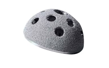
Acetabular prosthesis
The cementless acetabular prosthesis has several main characteristics:
Shape and Material: It is generally hemispherical in shape, with an outer shell (metal-back) often made of titanium alloy or high-nitrogen stainless steel, such as M30NW (ISO5832-9).
Bone fixation: The outer surface is designed to promote osseointegration, often with a porous coating (e.g., titanium plasma or HA/TPS) or specific texturing, ensuring long-term fixation.
Anchorage holes: It can have cluster holes or a specific number of holes (e.g., 1/3 holes) for the insertion of acetabular screws, improving initial stability.
Inner surface: The inner surface is polished to minimize wear of the joint between the insert and the cup.
Compatibility: It is designed to be combined with different types of inserts (e.g., polyethylene or ceramic) and femoral heads, allowing for great surgical flexibility.
Rotation and Positioning: Some models allow the component to rotate, facilitating optimal positioning in the recipient bone.

Orthopedic Bone Cutter for Spine Surgery
The instruments shown in the image are orthopedic rod cutters, also known as bone cutters or rod cutters for spinal surgery. Here are their main features:
Material: Made of high-quality stainless steel.
Cutting Diameter Range: Designed to cut rods or bones with a diameter ranging from 4.0 mm to 6.5 mm, providing high precision during operations.
Total Length: Approximately 550 mm, improving maneuverability during surgery.
Operation: Simple manual operation for precise cutting and increased patient safety.
Design: Ergonomically designed to reduce hand fatigue and ensure comfort during prolonged use.
Versatility: Suitable for a wide range of applications, particularly in orthopedic and spinal surgery.
Blades: Sharp blades for precise bone cutting, increasing surgical efficiency.
Specific Use: Can be used as a pin cutter for bone pins and for in situ cutting of 3.5 to 4.5 mm rods.

Instruments TLIF PEEK Cage
L’ensemble d’instruments TLIF PEEK Cage est un ensemble d’instruments chirurgicaux orthopédiques spécialisés, principalement utilisé pour la chirurgie de fusion intersomatique lombaire transforaminale (TLIF).
Caractéristiques principales :
Matériaux de haute qualité : Les instruments sont fabriqués à partir de matériaux durables, y compris l’acier inoxydable et le PEEK de qualité médicale (Polyétheréthercétone) pour les cages.
Précision et exactitude : Conçus pour permettre aux chirurgiens d’effectuer la procédure TLIF avec une grande précision.
Facilité d’utilisation : L’ensemble est conçu pour être convivial, simplifiant les étapes complexes de la chirurgie de fusion spinale.
Personnalisation : Offre une variété d’options d’instruments pour répondre aux besoins et préférences spécifiques des équipes chirurgicales.
Stérilisation : Les instruments sont conçus pour être facilement stérilisables, assurant l’hygiène et la sécurité lors des procédures chirurgicales.
Application : Principalement utilisé en chirurgie de la colonne vertébrale pour des conditions telles que la maladie dégénérative du disque, la sténose spinale et le spondylolisthésis.
Fonction de la cage TLIF PEEK : La cage PEEK est insérée dans l’espace discal après le retrait du disque blessé, visant à restaurer et maintenir la hauteur discale normale, à fournir une stabilité et à favoriser la fusion vertébrale. Elle est également radiotransparente pour un suivi postopératoire aisé.
Contenu de l’ensemble: Comprend généralement des instruments tels que des alésoirs, des forets, des curettes, des inséreurs d’implants, des écarteurs nerveux et des écarteurs d’espace discal.
