
Lens Cleaning Kit
The Gommax Lens Cleaning Kit features the following features:
Phosphate-Free Formula: Designed for safe and gentle lens cleaning.
Streak-Free Cleaning: Provides a streak-free finish for clear vision.
Suitable for All Lens Types: Safe to use on eyeglasses, sunglasses, and other optical surfaces, including coated lenses.
Complete Kit: Includes a lens cleaning spray and an ultra-soft microfiber cloth for effective cleaning.
Compact Size: The 1.7 fl oz (approximately 50 ml) spray bottle is convenient for on-the-go use.
Removes Dirt and Fingerprints: Effectively cleans dirt, grime, dust, and fingerprints.

Medical Adhesive Tape
The characteristics of medical adhesive tape, such as the “Adhesive Tape U.S.P.” shown in the image, are as follows:
Material and Adhesive: It is generally composed of a backing (often fabric, paper, or polyethylene) and an adhesive, which can be synthetic rubber or acrylic, formulated to adhere to the skin.
Hypoallergenic and Latex-Free: Medical tapes are often designed to be hypoallergenic and latex-free to minimize the risk of allergies and skin irritation, especially for sensitive skin.
Breathability: Many medical adhesive tapes are made with breathable materials, such as white paper, allowing the skin to breathe for increased comfort during extended use.
Adhesion and Strength: They provide sufficient adhesion to hold dressings or medical devices in place, yet are designed to be removed without causing excessive skin trauma.
Conformability: The flexibility of the backing allows the tape to conform to the contours of the body, ensuring long-lasting adhesion even on uneven surfaces.
Water Resistance: Some medical adhesive tapes are water-resistant to maintain their effectiveness even in the presence of moisture.

Lens Edger
Modèle :NAME: Lens Edger
The ALE-1000 Auto Lens Edger is a template-free lens edger with several notable features.
Key Features:
Screen and User Interface:
5.7-inch color TFT LCD screen with a high resolution of 640×480.
1:1 display of lens graphics, icons, and a user-friendly interface.
Design and Ergonomics:
Ergonomic design with a sleek shape.
Divided configuration between the main body and scanners.
Lens Processing Capabilities:
Supports various lens types, including CR-39 resin lenses, high-index lenses, super-hard coated lenses, glass lenses, PC lenses, and Trivex lenses.
Features two operating modes:
- optical center and geometric center.
- Automatic correction of scan data, including the axis and nose bridge distance.
- 100 mm diameter grinding wheel for processing highly curved lenses.
- Bevel polishing function for perfect lens fitting.
Scanning Features:
- High-precision, fast, and convenient 2D scanning.
- Ability to scan metal frames, plastic frames, demo lenses, and templates.
- Scanning options for one or both eyes, with automatic frame positioning and automatic calculation of the nose-to-eye distance.
- Automatic frame clamping and automatic determination of frame, liner, and template type.
Security and Stability:
Inherits the safety, stability, robustness, and durability of the LE series of lens edgers.

Optical Lens Cutting Machine
MODEL: SJG-1000
The features of the SJG-1000 (or SJG-1000AK) optical lens cutting machine are as follows:
Tracing and Scanning System:
Optical scanning and centering are positioned as a single system.
Obtain the frame shape in 0.5 seconds.
Real-time imaging guidance to confirm the optimal center position.
Automatic elimination of lens, prism, and artificial parallax effects.
Display and Interface:
7-inch high-resolution (800*600) color touch screen.
Simple operation and low failure rate, requiring little maintenance.
Lens Processing Capabilities:
Suitable for various lens materials, including glass, CR, PC, high-index lenses, and CR-39.
Equipped with various wheels (diamond for glass, V-groove for CR/PC, bevel polishing).
Processes lenses from 22 mm to 80 mm.
Automatic and manual beveling are available.
Grooving width, depth, and position are manually adjustable.
V-shaped and flat polishing are available.
Other Features:
Can be connected simultaneously to two SJG-1000AK automatic milling machines to form a processing center.
3D plotter for accurate lens imaging in 1 second on an 8-inch monitor (for the AK version).
Built-in, easy-to-use blocker (for the AK version).
Four-level adjustable scanning light source (for the AK version).
Compact design for easy placement in a workshop or laboratory.
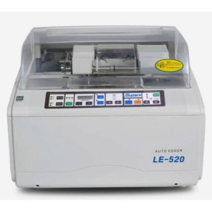
Auto Lens Edger
MODEL: LE-520
The LE-520 Auto Edger is an optical equipment designed for the automatic edger of eyeglass lenses.
Here are its main features:
Capable of processing CR39, glass, and PC (polycarbonate) lenses
Flat edge polishing and beveling functions
Equipped with a universal non-slip chuck
Standard PC functionality
Precision and Stability: Designed to process lenses with precision and stability
Ease of Use: Inherits the design style of Supore’s LE series, making it easy to use
Power Supply: 220V/50Hz
Unit Weight: Approx. 50 kg.
Power Consumption: 500W.
Technical Parameters (min/max):
Lens Size: Minimum 22 mm, Maximum 100 mm.
Adjustable Measuring Range: -6.0 mm to +6.0 mm in 0.05 mm increments.
Dimensions: 510 mm x 490 mm x 40 mm.
Grinding Wheels (Typical Configuration): Includes wheels for glass, CR, and polycarbonate, as well as a polishing wheel and a V-groove wheel.

Non-Contact Tonometer
The NCT-100 non-contact tonometer is an ophthalmic device designed for measuring intraocular pressure (IOP) using a puff of air.
Its main features are:
Fully Automatic Measurement: It allows for fully automatic measurement of data for both eyes without the need for manual switching.
Automatic 3D Positioning: Automatic 3D (xyz) positioning ensures accurate and reliable measurements.
Ease of Use: The device is easy to use thanks to its color touchscreen.
Data Stability: It provides stable IOP data thanks to a gentle puff of air measurement.
Corneal Thickness Compensation: It is capable of compensating for corneal thickness during IOP measurement.
Speed: It provides measurement results in less than 10 seconds.
Non-contact: The non-contact measurement method reduces the risk of eye injury or infection.

Ophthalmic Operating Table
The features of an ophthalmic operating table, such as the one shown, generally include the following:
Specialized Design for Ophthalmology: The table is specifically designed for ophthalmic surgeries and treatments, providing optimal positioning for delicate ocular procedures.
Triple-Section Tabletop: The tabletop is often divided into three sections, with a detachable head section and an integrated drainage system to facilitate eye surgeries.
Durable and Easy-to-Clean Materials: The base and upholstery are made of stainless steel, ensuring good stability, corrosion resistance, and easy cleaning, thus reducing the risk of infection.
Ergonomic Adjustments: The table allows for various position adjustments (height, Trendelenburg, lateral tilt) for patient comfort and surgeon ergonomics, often controlled by a manual unit.
Special Accessories: It is equipped with an ophthalmic headrest for stable positioning of the patient’s head and can include padded arm supports for the surgeon.
Operation: Available in hydraulic or electric options, providing smooth and precise tabletop movement for precise patient positioning.

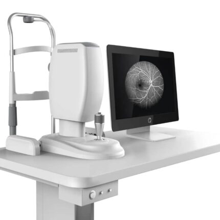
Optical Coherence Tomograph
The device shown is a Nidek RS-3000 Advance 2 optical coherence tomograph (OCT). Its key features include:
Comprehensive Retinal and Glaucoma Analysis: It offers a complete solution for analyzing retinal pathologies and glaucoma.
SLO-Based Eye Tracker: A scanning laser ophthalmoscope (SLO)-based eye tracker system enables precise image capture with eye movement compensation.
Selectable OCT Sensitivity: Enables B-scan image acquisition even through opacities in the ocular media.
HD Plus Tracing: This feature ensures accurate averaging of up to 120 images for high-resolution, high-contrast images.
OCT Angiography (OCT-A): High-resolution OCT angiographic imaging capability for observing microvascular details of the retina.
Glaucoma Analysis with Normative Database: Includes glaucoma analysis with an extensive normative database (9 x 9 mm).
High Scanning Speed: The device can reach 85,000 A-scans per second, enabling rapid image acquisition.
Automatic Panoramic Imaging: Automatic panoramic imaging capability for OCT angiography up to 12 x 12 mm.
Vibrant Color LCD Display: Equipped with an 8.4-inch color LCD display for real-time data viewing and review.
Comfortable Working Distance: A 35.5 mm working distance ensures comfortable patient positioning.
Color Fundus Camera
The device shown is a color fundus camera, a confocal ophthalmoscope for retinal angiography.
Its key features include:
Confocal laser scanning technology: Enables high-resolution imaging and stereoscopic viewing for depth perception.
High-resolution video capture: Ability to record videos at high resolutions, up to 1024 x 1024 pixels.
Multi-band fluorescence imaging and photography: Allows the use of different dyes such as fluorescein and indocyanine green to visualize retinal and choroidal vessels, although OCT angiography can reduce the need for dyes.
Non-mydriatic and continuous operation: Ability to capture images without prior pupil dilation, providing smoother operation.
Wide Field of View: Capable of capturing a 65-degree angle in a single undilated fundus photograph, with composite fields of view up to 120°.
High Resolution: Up to 1536 x 1536 pixels for high-resolution images.
Multiple Laser Sources: Uses different laser wavelengths (e.g., 488 nm for FA, 795 nm for ICGA, 830 nm for IR) for various imaging applications.

Ophthalmic YAG Laser System
MODEL: MD-920
The MD-920 ophthalmic YAG laser system, as shown in the image, has the following specifications:
Laser Specifications:
Laser Type: Nd:YAG, Q-switched
Wavelength: 1064 nm
Mode: Q-switched (multimode)
Pulse Output: Single pulse, double pulse, and triple pulse burst
Pulse Duration: 4.5 ns
Repetition Rate: 2.5 Hz
Cone Angle: 18°
Spot Size: 30 µm
Aiming Beam Wavelength: 633 nm
Aiming Beam Output Power: <0.4 mW
Slit Lamp (Galilean Stereoscopic Microscope) Features:
Provides high clarity and sharpness, avoiding double images.
Five-level drum magnification.
Optional accessories: teaching tube, CCD camera, and tonometer.
Other Features:
Easy setup for installation and maintenance.
Mounted on a mobile rolling stand for transport.
Uses microprocessor control technology with a digital display for easy operation.
Built-in laser protection system to ensure surgical safety.

Matronix B-scan Biometer
The Matronix B-scan Biometer is an advanced ophthalmic ultrasound system designed for the diagnosis and monitoring of ocular conditions. Its key features include:
High-resolution imaging: Provides clear and detailed images of internal ocular structures, with 256-level grayscale imaging for exceptional tissue differentiation.
Probes: Typically equipped with a 10 MHz probe for accurate measurements.
Display: Incorporates a large 10.4-inch or 12.1-inch LED display for comfortable viewing and easy image interpretation.
Scanning modes: Combines A-scan and B-scan modes, allowing for distance measurements (ACD, lens, vitreous, AXL) and observation of intraocular and posterior segment diseases.
Portability and Design: Compact and portable tabletop model (dimensions: 256 x 150 x 326 mm; weight: 4.5 kg), ideal for clinics and mobile environments.
Power Supply: Mains powered (AC 100-240V) and often equipped with a 4400 mAh lithium battery for stand-alone use.
Additional Features: Supports single and multiple image selection, real-time dynamic scanning to display lesion location, volume, and shape, and can be connected to a video recorder or ultrasound workstation.
Clinical Applications: Used for the diagnosis of ocular and orbital diseases, including refractive media turbidity, ocular somatometry, pre- and post-vitrectomy diagnosis, intraocular foreign body localization, intraocular tumors, exophthalmos, and ocular and orbital hemodynamics.

Phacoemulsifier
MODEL: MD-480A
The Meda MD-480A Phacoemulsifier features:
Control and Monitoring: All parts of the system are fully controlled and monitored by microprocessor technology.
Ultrasonic Emulsification (U/S):
Phaco Emulsification Frequency: 40 kHz ± 20%.
U/S Handpiece Upper Tip Vibration Accuracy: Max. 120 μm ± 20%.
Operating Modes: Continuous, Pulse, Linear, Burst.
Emulsification Power: 0-100%.
Pulse Frequency: 1 Hz to 99 Hz.
Burst Time: 5 ms to 100 ms.
Irrigation/Aspiration (I/A):
Peristaltic pump for aspiration.
Maximum flow rate: 40 ml/min.
Reflux flow rate: 20 ml/min.
Maximum vacuum: 66.7 kPa (500 mmHg).
Vitrectomy:
Cutting frequency: 20 to 600 cuts/min.
Cutting power source: Air pump.
Cauterization:
Frequency: 1 MHz.
Maximum power: 10 watts (with a 200 Ω load).
Power range: 0-100%.
User interface: Large TFT touchscreen and user-friendly menu.
Additional features:
Up to 10 sets of preset parameters.
Automatic testing for tubing and handpiece problems.
Intelligent voice reminder.
Electrical Requirements: 220 Volts ± 10%, 50 Hz ± 2%.
Dimensions and Weight: 43 cm (W) × 22.5 cm (H) × 32 cm (D), Weight: 8 kg.
