
Hand- held fundus cameraV
Portability & Compactness: Designed to be ultra-lightweight and compact, allowing for easy handling and use in diverse settings like community and low-resource environments.
Non-mydriatic Operation: Many devices operate without dilating the pupils, making the exam more comfortable for patients and efficient for healthcare providers.
Ease of Use: Features such as auto-focus, auto-exposure, and an on-screen targeting aid (internal fixation LEDs) simplify the image acquisition process, even for less experienced users.
Connectivity: Support wireless and wired data transfer (WLAN, USB) and cloud integration for image management, remote analysis, and integration into hospital systems.
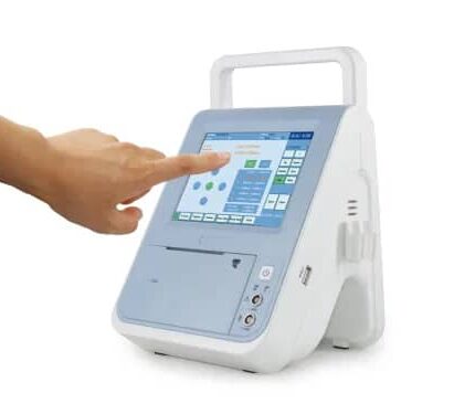
Scan and Pachymeter
Features:
- Excellent quality, Compact design, Defect free, and Axial Length Measurement 10MHz Probe with Fixation Light
- The Occuity PM1 pachymeter will make it easier, faster and safer to diagnose glaucoma by measuring corneal thickness in just a few seconds.
- Automatic reading at single or multiple points for corneal thickness; Multiple measurements at single point for higher reliability; Higher accuracy enabled by ..
- Precise Measurement with both Immersion & Contact modes. Touch Screen Extremely Easy of Use Compact and Portable Design Auto/Manual .
Specifications: A scan probe: 10MHz import small size probe, built-in luminotron. Measuring range: 15mm-40mm. Measurement precision: ±0. 05 …
A-Scan Function: Measures the axial length of the eye and the thickness of internal structures like the lens and retina. This is crucial for accurately calculating the power of intraocular lenses (IOLs) before cataract surgery.
Pachymeter Function: Measures central corneal thickness (CCT) by detecting reflections from the cornea’s anterior and posterior surfaces.
Technology: Can use either ultrasonic waves or focused light to take measurements.
Measurement Types: Offers both immersion and contact methods for A-scan and ultrasound for pachymetry.
Accuracy & Precision: Provides highly accurate and precise measurements, often with electronic resolutions and accuracies of 0.1 mm or better.
Operating Modes: Features automatic and manual capture modes for user preference.
Data Handling: Includes capabilities for storing patient data, EMR/EHR compatibility, and network connectivity.

Scan and Pachymeter SW1000
Functions: Measures axial length, anterior chamber depth, and lens thickness.
Macular Recognition: A built-in function for accurate measurement of axial length.
IOL Calculation: Includes formulas like SRK-II, SRK-T, Hoffer-Q, Holladay, Binkhorst-II, and Haigis.
Eye Modes: Supports phakic, aphakic, and dense eye measurements.
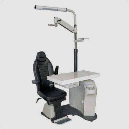
Complete Workstation Designed For Eye Examinations
The ophthalmic unit illustrated is a complete workstation designed for eye examinations, incorporating several key components:
Examination chair: A height-adjustable, often electrically powered, swiveling seat, providing comfort for the patient and flexibility for the ophthalmologist.
Instrument table: A lifting, sometimes sliding, table designed to accommodate various ophthalmic instruments such as a refraction head, an autorefractor, or a slit lamp.
Lighted column and supports: A vertical column incorporating an examination light (often halogen with adjustable lighting) and supports for other equipment, such as a chart projector or a motorized phoropter arm.
Control panel: Integrated controls on the table or a footswitch for adjusting the chair height and other unit functions.
Built-in storage: The unit often includes a cabinet or drawers for storing trial lenses and other small accessories.
Power supply: Built-in power transformers to power the instruments, as well as outlets for additional devices.
Ergonomic and compact design: Many units are designed to optimize space and facilitate intensive use in medical settings, with particular attention to stability and robustness.
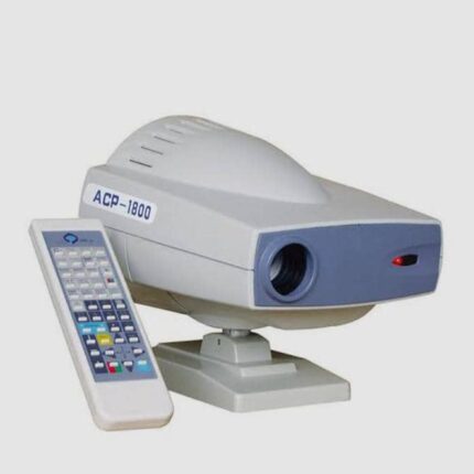
Automatic Chart Projector
MODEL: ACP-1800
The ACP-1800 Automatic Chart Projector is an ophthalmic instrument used for vision testing. Its main features are:
Projection Distance: It can project images at distances ranging from 1.5 m to 6 m.
Projection Magnification: The magnification is 30x at a distance of 5 m.
Projection Size: The projected image size is 330 mm (width) x 270 mm (height) at 5 m.
Number of Charts: It has 30 types of charts or graphs for vision testing.
Chart/Mask Change: Card or mask change is fast, taking 0.03 seconds per chart or mask image.
Lamps: It uses a 12V, 50W halogen lamp.
Programmability: Icons can be displayed in a predefined sequence via programming, and a single icon can be displayed via a multi-function mask plate.
Easy Installation and Maintenance: Bulb replacement is simple and requires no adjustment of focus or position.
Automatic Shutdown: The device automatically turns off after 10 minutes of inactivity.
Accessories: It comes with a remote control, a polarized metal shield, polarized glasses, fuses, and batteries.
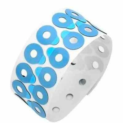
Adhesive tape
For Lens Processing:
Adhesion: Must securely bond with the blocking wax or alloy used for holding the lens.
Protection: Prevents scratches and damage to the lens surface during grinding and polishing.
Peelability: Must peel off cleanly and easily from the lens after processing, without leaving residue or transferring alloy.
Color: Often blue to make it easy to distinguish and remove from the lens.
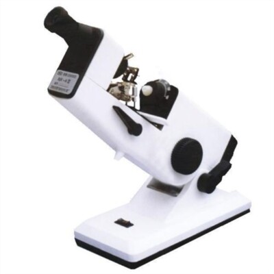
Hand lens meter
Lens power Measurement: Can measure spherical power, cylindrical power, and axis (direction) of cylindrical lenses.
Prism Power & Base Direction: Determines the strength of a prism and its base direction.
Optical Center Marking: Includes a precision inking attachment to mark the lens’s optical center for cutting and edging.
Used for quality control and manufacturing of lenses.
Opticians’ Shops: Essential for verifying prescription lenses and fitting glasses.
Ophthalmology: Employed in hospitals and clinics for comprehensive eye.
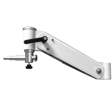
Electric chair/ Phoropter arm
Motorized Operation: Uses an electric mechanism for effortless and precise horizontal (and sometimes vertical) adjustments, reducing physical on the clinician.
Smooth and Stable Movement: Provides stable, controlled positioning of the phoropter, which is crucial for accurate ophthalmic measurements.
Durability: Constructed from high-quality, durable materials to ensure long-lasting performance in a clinical setting.
Precision Adjustment: Allows for fine-tuned, accurate positioning of the phoropter, improving the efficiency and precision of refractions.
Clinician Convenience: Designed for easy installation on compatible stands and examination units, improving workflow in a modern practice
Patient Comfort: Contributes to patient comfort by enabling smooth and effortless patient positioning.

Leather Gynecological Apron
This waterproof PVC gynecological apron features the following features:
Material: Made of durable PVC (polyvinyl chloride).
Waterproofness: Provides excellent resistance to water, blood, chemicals, and oils, making it ideal for humid work or retail environments.
Protection: Designed to cover the entire body, it provides effective protection against splashes and dirt.
Ease of Maintenance: Easy to clean and disinfect after use.
Comfort: Lightweight and easy to wear for extended periods of work.
Design: Includes adjustable neck and waist straps for a secure and comfortable fit.
Color: Available in bright colors like orange for better visibility and identification.

Reinforced Gown
The disposable reinforced gown has several key features:
Material and Use:
Made of polypropylene (PP) or polyethylene (PE) non-woven fabric, providing protection against splashes and contamination.
Designed for single use to prevent cross-contamination.
Design and Protection:
Features long sleeves for extended arm protection.
Can include elastic or tight cuffs for a secure fit and enhanced protection.
The design can be breathable for wearer comfort, while maintaining fluid barrier properties.
Functionality:
Easy to put on and take off, with ties at the waist and neck for a customizable fit.
Used in a variety of environments, including hospitals, laboratories, or for visitors in hazardous areas, to protect both the wearer and the environment.
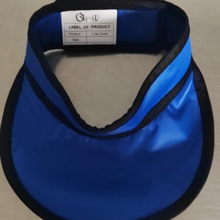
X-ray Protective Collar
The X-ray protective collar, also known as a lead thyroid collar, has the following features:
Radiation Protection:
It is designed to provide optimal protection of the neck and thyroid area from X-rays during radiological examinations. It has a lead equivalent of 0.3 mm or 0.5 mmpb for effective protection.
Design and Materials:
Made of durable lead rubber or polyurethane-coated fabric.
Ease of Use:
The Velcro design on the back allows for easy on and off and allows the collar to be adjusted for a comfortable and secure fit.
It is designed to be lightweight and soft for the comfort of the patient or professional.
Durability:
It is robust and designed for durability, ensuring extended use.
Applications:
Suitable for various medical settings, including dentistry, hospitals, and X-ray offices.

Operating Room Pajamas
The characteristics of the operating room gown, which is a disposable operating room pajama, are as follows:
Materials: Generally made of latex-free, hypoallergenic non-woven polypropylene. Cotton versions are also available, offering lightness and breathability.
Weight: Around 40 g/m² for non-woven versions.
Color: Most often blue, as shown in the image.
Comfort and Hygiene: Designed to provide comfort and ease of movement for hospital staff, while meeting the hygiene standards required in surgical environments.
Use: Mainly used during surgical procedures by hospital staff.
