
Birthing Pelvis with 2 Babies (Male and Female)
The Birthing Pelvis: Structure and Function in Childbirth
The female birthing pelvis serves as the crucial bony framework that enables successful vaginal delivery. This specialized structure combines anatomical precision with dynamic adaptability to facilitate the birth process.
Bony Architecture:
-
Composed of four interconnected bones:
-
Paired ilium bones (forming the sides)
-
Sacrum (posterior wall)
-
Coccyx (tailbone)
-
-
Forms a complete pelvic ring connecting the spine to the femurs
-
Provides structural support while allowing necessary movement
Morphological Features:
-
Distinctive hourglass configuration with:
-
Expanded false pelvis (greater pelvis) superiorly
-
Constricted true pelvis (lesser pelvis) inferiorly
-
Demarcated by the pelvic brim (superior strait)
-
-
Exhibits characteristic female adaptations:
-
Wider subpubic angle (>90°)
-
Broader, more circular inlet
-
Shorter sacral curvature
-
Obstetric Dimensions:
Three critical planes determine fetal passage:
-
Pelvic Inlet (Superior Strait):
-
Key diameter: Promontoretropubic (11.5-13 cm)
-
-
Midpelvis:
-
Crucial measurement: Median transverse (10.5 cm)
-
-
Pelvic Outlet:
-
Important span: Interspinous (10-11 cm)
-
Dynamic Adaptations:
-
Hormonally-mediated changes (relaxin, progesterone):
-
Increased sacroiliac joint mobility
-
Pubic symphysis widening (2-3 mm)
-
Ligamentous relaxation
-
-
Permits up to 10-15% expansion of birth canal dimensions
Parturition Mechanics:
-
Fetal Engagement: Head enters pelvic inlet
-
Descent: Progressive movement through planes
-
Rotation: Internal adjustment to navigate contours
-
Expulsion: Final passage through outlet
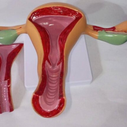
Healthy Uterus
Features of a healthy uterus model, such as this one, include the following:
Detailed Anatomical Representation: The model shows the three-layered structure of the uterus (endometrium, myometrium), the uterine cavity, the cervix, and the vagina, often with the fornix visible.
Associated Female Reproductive Organs:
It includes the ovaries and fallopian tubes (oviducts), illustrating their relationship to the uterus. Sections may show follicles and the egg in various stages of maturity.
Open Vagina and Cervix:
The vagina and cervix are cut longitudinally to allow internal visualization of the uterine lining and uterine muscles.
Life-Size and Realistic:
The model is generally life-size for better anatomical understanding, with hand-painted structures for increased realism.
Educational Use:
Designed as an educational tool for teaching female anatomy, it is useful for medical students and healthcare professionals.

Pregnant Female Anatomy
An anatomical model or medical simulator, likely used for teaching or training in obstetrics or gynecology.
Its main features are as follows:
Anatomical Representation:
This is a detailed model of the pregnant uterus, showing the position of the fetus inside.
Materials:
Made from soft, realistic materials to simulate human tissue.
Functionality:
Designed to simulate obstetric procedures or gynecological examinations, allowing students to practice palpation, simulated deliveries, or other procedures.
Visible Details:
The model allows for observation of the relationship between the fetus and the uterus, as well as the structure of the pelvis.
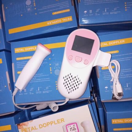
Fetal Doppler
his professional-grade fetal doppler is designed for accurate, safe, and convenient fetal heart monitoring with the following features:
1. High-Performance Probe
-
High sensitivity for early detection (typically from 12 weeks)
-
Precision accuracy (±1 BPM) in heart rate measurement
-
Ergonomic design for comfortable handling
2. Superior Audio Quality
-
Adjustable volume control with high-fidelity sound output
-
Noise reduction technology for clear fetal heartbeat detection
-
Dual audio modes:
-
Built-in speaker for shared listening
-
Earphone jack for private monitoring
-
3. User-Friendly Display
-
Backlit LCD screen for clear visibility in all lighting conditions
-
Real-time fetal heart rate (FHR) display
-
Battery level indicator
4. Safe & Hygienic Design
-
Radiation-free operation (ultrasound power <10mW/cm²)
-
Medical-grade ABS plastic construction:
-
Durable & impact-resistant
-
Easy to clean & disinfect
-
5. Power & Portability
-
Rechargeable lithium battery (4-6 hours continuous use)
-
USB charging for convenience
-
Compact & lightweight for clinic or home use

Optical Lens Trial Case
The optical lens trial case has the following characteristics:
Type of Equipment:
This is a trial lens set (or “Trial Lens Set”), used in optometry and ophthalmology.
Contents:
It contains a full range of spherical lenses (positive and negative), cylindrical lenses (positive and negative), and prisms, as well as accessories such as a trial frame and an occluder.
Use:
This set is essential for determining the necessary visual correction during an eye exam, allowing you to test different lens powers to refine your optical prescription.
Case Materials:
The case is rigid, made of aluminum or a similar material, with a padded interior to protect the lenses.
Organization:
Lenses are generally systematically organized by dioptric power and type (spherical, cylindrical) to facilitate quick selection by the practitioner.
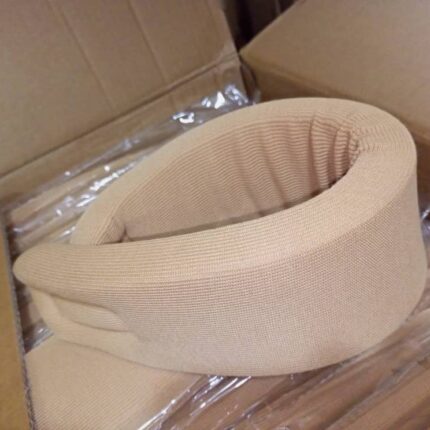
Cervical Collar
Navid Code 154 Medical Cervical Collar – Features & Applications
Key Specifications:
-
Materials: Soft sponge core with breathable fabric cover
-
Closure System: Adjustable hook-and-loop (Velcro®) strap for secure fit
-
Hypoallergenic: Anti-irritation design for sensitive skin
-
Origin: Manufactured in Iran
Design & Comfort:
-
Balanced Support: Maintains natural cervical alignment while allowing gentle mobility
-
Lightweight & Comfortable: Ideal for prolonged wear without skin irritation
-
Contoured Shape: Anatomically designed to reduce pressure on the jaw and occiput
Medical Applications:
-
Cervical Stabilization:
-
Post-traumatic support (whiplash, minor cervical injuries)
-
Management of cervical osteoarthritis (spondylosis)
-
Torticollis (congenital or acquired) correction
-
-
Therapeutic Benefits:
-
Reduces nerve root compression via mild axial traction
-
Alleviates muscle spasms and ligament strain
-
Prevents abnormal neck flexion/extension
-
-
Post-Surgical Use:
-
Supplemental support after cervical spine procedures
-
Target Conditions:
✔ Cervical radiculopathy
✔ Cervicalgia (neck pain syndromes)
✔ Mild cervical disc herniation
✔ Postural neck deformities
Advantages:
-
Non-restrictive for daily activities
-
Adjustable fit accommodates varying neck circumferences
-
Easy to clean (hand wash recommended)
Note: For moderate to severe instability, consult a physician for rigid collar alternatives.

Ear Piercing Gun
Professional Ear Piercing Gun – Features & Specifications
Complete Piercing Kit Includes:
✔ Piercing gun – Precision-engineered for accurate placement
✔ Medical-grade marking pen – For symmetrical dot placement
✔ Compact storage case – Hygienic and portable
✔ Round inspection mirror – Allows for self-application
Premium Construction:
-
High-grade stainless steel & metal alloy – Durable, corrosion-resistant, and long-lasting
-
Ergonomic grip – Ensures steady control during piercing
User-Friendly Design:
-
Quick-release mechanism – Enables fast, single-action piercing
-
Adjustable tension control – For varying ear cartilage thickness
-
Lightweight & portable – Ideal for professional studios, clinics, or home use
Hygiene & Safety Features:
-
Autoclavable (or disposable cartridge options) – Reduces cross-contamination risks
-
Pre-sterilized hypoallergenic studs – Minimizes infection risk
-
Single-use safety lock – Prevents accidental re-use
Versatile Applications:
-
Primary Use: Standard earlobe piercing
-
Adaptable For:
-
Helix/cartilage piercings (with proper attachments)
-
Nose stud application (select models)
-
Navel piercing (specialized variants)
-
Ideal For:
-
Professional piercers – Ensures consistent, sterile results
-
Clinics & dermatologists – Medical-grade safety for patients
-
Home users – Easy self-piercing with proper precautions
Note: Always follow sterilization protocols and aftercare instructions to prevent complications.
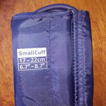
Pediatric Cuff
Pediatric Blood Pressure Cuff – Features & Specifications
Key Attributes:
✔ Small Cuff Design – Optimized for young patients
✔ Arm Circumference Range: 17–22 cm (6.7″–8.7″)
✔ Universal Compatibility – Fits most standard BP monitors via interchangeable connectors (4.6 mm, 5 mm, 6 mm, 7 mm)
Material Composition:
-
Outer Layer: High-density nylon for durability & easy cleaning
-
Inflatable Bladder: Medical-grade non-toxic TPU for safety & accuracy
-
Hook-and-Loop Closure: Secure yet gentle on delicate skin
Clinical & Practical Benefits:
-
Accurate Measurement: Ensures proper fit to avoid false high/low readings
-
Adjustable Fit: Contoured shape prevents slippage during inflation
-
Comfort-Focused: Soft edges minimize discomfort for children
Intended Use:
-
Routine pediatric BP monitoring (hospitals, clinics, home use)
-
Ideal for toddlers to school-aged children
-
Compatible with manual & digital sphygmomanometers

ECG Electrode
Disposable ECG electrodes have the following characteristics:
Product Type: Disposable ECG Electrodes.
Sensor: Ag/AgCl sensor (Silver/Silver Chloride).
Packaging: Each pack contains 50 pieces.
Compliance: CE marked, indicating compliance with European standards.
Use: Designed for electrocardiography (ECG).
Gel: Generally equipped with a conductive gel for good signal transmission.
Backing: Made with a foam or non-woven backing for patient comfort and good adhesion.

Ultrasound Printer Paper
Sony UPP-110HG High Gloss Thermal Paper – Technical Specifications
Media Characteristics
-
Type: V-Class High Gloss Thermal Film
-
Finish: Premium glossy surface (25% brighter than UPP-110HD)
-
Water Resistance: Splash-proof coating for image durability
Physical Dimensions
-
Roll Width: 110 mm
-
Roll Length: 18 meters
-
Print Capacity: ±200–240 A6 prints (depends on margin settings)
Printer Compatibility
Supported Sony B/W Thermal Printers:
✔ UP-897MD | UP-D895/D897/D898MD | UP-X898MD
✔ UP-895CE/897CE | UP-D890/D890 CE
✔ UP-898 | UP-D898 | UPX-898MD
✔ UP-897 | UP-D897 | UP-890/D890
✔ P-860 | UP-D860
Image Performance
-
Optical Density: High-contrast B/W output
-
Resolution: Sharp detail reproduction
-
Gloss Level: 85°+ (measured at 60° gloss meter)
Primary Applications
-
Medical Imaging:
-
Obstetric/Gynecological ultrasounds
-
Urological diagnostics
-
Radiological scans
-
-
Clinical Documentation:
-
Ultrasound snapshots
-
Procedure records
-
Environmental & Handling
-
Storage Conditions: 10–25°C, <70% humidity
-
Shelf Life: 2 years (unopened, in original packaging)
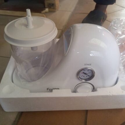
Single-jar electric aspirator
Electric Aspirator – Model HOO3-A
Product Overview
The HOO3-A single-jar electric aspirator is a medical-grade suction device designed for efficient fluid and mucus extraction in clinical and home-care settings.
Key Features & Specifications
General Characteristics
-
Type: Electric suction unit (portable/tabletop)
-
Applications:
-
Oral/nasal/tracheal suctioning
-
Postoperative care
-
Tracheostomy management
-
Minor surgical procedures
-
-
Patient Suitability: Adults & pediatric use
Core Components
✔ Collection Jar
-
Material: Autoclavable polycarbonate
-
Capacity: 1000 mL (1L)
✔ Vacuum System
-
Adjustable suction power (via regulator)
-
Pressure gauge (displays kPa/bar)
-
Max. negative pressure: >0.075 MPa (~563 mmHg)
-
Flow rate: 16 L/min (model-dependent)
✔ Hygiene & Maintenance
-
Autoclavable parts: Jar, tubing
-
Disposable components: Antibacterial filters, suction catheters
Technical Data
-
Power Supply: AC 220V, 50Hz
-
Power Consumption: 250VA
-
Noise Level: <50 dB (quiet operation)
Intended Use Environments
-
Hospitals & clinics
-
Home healthcare
-
Emergency care units

Hematology Machine
URIT-3000Plus Hematology Analyzer – Technical Specifications
Key Features
✅ Fully Automated 3-Part Differential Analyzer
✅ High Throughput: Up to 60 samples/hour
✅ 21 Measured Parameters + 3 Histograms
Technical Specifications
Analysis Principles
-
Electrical Impedance: WBC, RBC, PLT counts
-
Photoelectric Colorimetry: HGB measurement
Performance & Efficiency
-
Sample Volume: 20 μL (capillary or venous blood)
-
Memory Capacity: 100,000+ results (with histograms)
-
Data Export: USB, HL7-compatible (LIS/HIS integration)
User Interface & Operation
-
10.4-inch Color LCD (Linux OS)
-
Input Options:
-
Standard keyboard & mouse
-
Optional touchscreen upgrade
-
-
Real-time monitoring of system status
Quality & Compliance
-
Certifications: CE, ISO 13485
-
Built-in QC: Supports 3-level calibration
Maintenance & Service
-
Self-Check Diagnostics: Minimizes downtime
-
Low-Consumable Design: Cost-effective operation
