
Hot Water Bottle
The features of a hot water bottle generally include:
Material: Often made of natural rubber or silicone, materials known for their heat retention and durability.
Capacity: Hot water bottles are available in various capacities, with 2 liters being a common size, ideal for extended use or for warming a larger area.
Leak-Proof Design: They feature a wide opening and a plastic screw cap to ensure a perfect seal and minimize the risk of leaks or burns.
Heat Retention: The material design and sometimes a ribbed outer surface help retain heat longer, reducing the need for refills.
Versatility: Hot water bottles are used not only for heat therapy (relief from muscle pain and cramps) but also, with the appropriate accessories, for enemas or showers, offering dual functionality.

Human Brain Anatomical Model
The human brain anatomical model has the following features:
Material: Made of high-quality PVC, ensuring durability and ease of cleaning.
Dimensions and Scale: Life-size (1:1 scale) for an accurate representation of the size and shape of the human brain.
Detachable Components: Composed of several parts, which can be disassembled for a detailed study of the various brain structures.
Structure Representation: Highlights various parts of the brain such as the cerebral hemispheres, lobes (temporal, occipital), brainstem, cerebellum, diencephalon, midbrain, pons, medulla oblongata, and cranial nerves.
Functional Area Identification: Some models use contrasting colors and labels to identify motor and sensory centers, as well as specific functional areas of the brain (limbic system, emotional centers, etc.).
Educational Use: Designed as a teaching tool for schools, hospitals, and physical health training, facilitating the understanding of brain anatomy.
Display Stand: Comes with a display base for stable and convenient presentation.

Human Digestive System Model
Features of the Human Digestive System Model
The human digestive system model presented, such as the Axis Scientific model, has several distinctive features.
Complete and Detailed Representation: It depicts the entire digestive system, from the mouth to the rectum, including key organs such as the esophagus, stomach, liver with gallbladder, pancreas, spleen, and intestines (small and colon).
Size and Scale: It is life-size.
High-Quality Material: Made of PVC, making it durable, resistant to high temperatures, and non-deformable.
Open and/or Removable Organs: Sections such as the duodenum, cecum, and rectum are open for better internal visualization. Parts such as the transverse colon or the anterior stomach wall are removable for further study.
Anatomically Accurate: Designed to be anatomically correct, developed by medical professionals, and hand-painted with meticulous attention to detail for enhanced realism.
Teaching Tool: Ideal for teaching anatomy and physiology in classrooms, clinics, or patient education, sometimes with numerical labeling to facilitate structure identification.
Stand Base: Mounted on a base or board for easy display and study, suitable for tabletop or wall mounting.
Accompanying Manual: Often comes with a detailed, full-color manual that identifies the various parts and provides educational information.
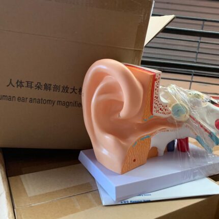
Human Ear Anatomical Model
A detailed, magnified model of the human ear designed for educational purposes in medical and academic settings.
Main Features:
- 5x Magnification: Enlarged five times the size of a real human ear for clear visualization of internal and external structures
- Detachable Design: Disassembles into at least two main parts (middle and inner ear) to facilitate study of overlapping elements
- Sturdy Material: Constructed from durable, wear-resistant PVC plastic with an ABS base for enhanced stability during display or teaching
- Labeled Structures: Precisely labeled parts, including outer ear, middle ear, inner ear, and cochlea, with color-coded sections for easy identification
- Versatile Use: Ideal for educational settings such as schools, hospitals, and laboratories, suitable for students, healthcare professionals, and educators

Human Hand Anatomical Models
Human hand anatomical models possess several important features for the study of anatomy:
Detailed representation of structures: They illustrate the bones, muscles, ligaments, nerves, and arteries of the human hand, providing a comprehensive view of the anatomy.
Durable Materials: Made of durable PVC, these models are designed to be robust and durable, withstanding frequent use in educational settings.
muscles
Anatomical Accuracy: Developed by medical professionals, these models are designed to be 100% anatomically correct, making them ideal for teaching and learning.
Support Base: Most models are mounted on a stable base, facilitating observation and manipulation during study.
Educational Uses: They are valuable tools for medical training, classroom teaching, and improving patient communication, particularly when explaining conditions such as arthritis.

Human Hand Model
Characteristics of the Human Hand Model:
The human hand model has several distinctive features.
Material: It is generally made of PVC plastic, making it odorless, durable, environmentally friendly, and portable.
Anatomical Details: The model faithfully reproduces the bony structure of the hand, including the eight carpal bones, five metacarpal bones, and fourteen phalanges.
Flexible Joints: Each joint is fixed with high-quality stainless steel wire, providing limited flexibility to simulate the natural movement of the hand.
Scientific Accuracy: This is a life-size, scientifically accurate hand skeleton, making it an excellent teaching tool for studying anatomy.
Standing Base: The model is mounted on a lightweight white base, making it easy to display and transport for teaching or demonstration.
Application: Designed for educational and demonstration purposes, it is useful for medical students, practitioners, and for display in classrooms or medical environments.

Human Leg Muscle Anatomical Model
The 13-part human leg muscle anatomical model features:
Detailed Representation: The life-size model shows the human leg musculature in detail, including superficial and deep muscle structures, vessels, nerves, and ligaments.
Anatomical Components: It represents the foot, the lower and upper leg, and half of the pelvis.
Removable Parts: The model includes 13 removable muscle parts for further study, such as the tensor fasciae latae, the rectus femoris, the extensor digitorum longus, the plantar fascia, the semitendinosus and semimembranosus muscles, the gluteus medius, the soleus, the gracilis, the gastrocnemius, the gluteus maximus, the long head of the biceps femoris, and the sartorius.
Anatomical Accuracy: Designed with high anatomical accuracy, it is a reliable resource for learning and examining the muscles of the human leg.
Identifiable Structures: It includes 64 identifiable structures, providing a thorough understanding of the muscular anatomy of the leg.
Materials: Made with high-quality materials to ensure longevity.
Dimensions and Weight: The model measures 103 x 19 x 17 cm and weighs 9.7 kg. It comes with a stand and manual.

Human Skull Anatomical Model
Human Skull Model Features
This human skull model features several notable features, ideal for anatomical study:
Life-Size: The model is a faithful reproduction of the life-size human skull, allowing for realistic visualization of proportions.
Disassemblable Structure: It is designed to be disassembled into several parts, such as the cranial vault, skull base, and lower jaw, facilitating the study of internal structures and bone relationships.
Anatomical Details: The model reproduces important anatomical details, including the cranial sutures and the dentition, which is similar to real teeth in terms of positioning.
Materials and Finish: Made of PVC plastic, it represents a natural bone color for a realistic appearance.
Articulated Jaw: The lower jaw is articulated and removable, allowing for the simulation of chewing movements.

Human Trachea Intubation Model
Features
1. Accurate, full-fledged anatomy of the mouth, tongue, airway, and esophagus. Perfect for moving endotracheal tubes.
2. Respiratory auscultation.
3. Oral and nasal intubation.
4. Oral and nasal suction.
5. Soft neck with crocodile cartilage allows for classic Sellick maneuvering, necessary to provide a better view of the vocal cord.
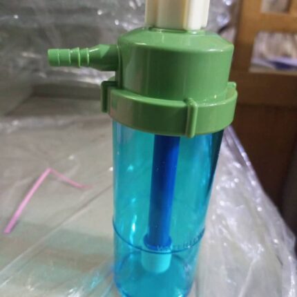
Humidifier
An oxygen humidifier is used to humidify oxygen before it is inhaled by the patient.
Its main features are as follows:
Function:
It provides moisture to dry oxygen from an oxygen source (such as a concentrator or oxygen cylinder) using a bubble-type humidification process.
Composition:
It is made from materials such as PC (polycarbonate) or polypropylene for the canister, and includes chrome-plated metal fittings.
Capacity:
The typical capacity of such a canister is 170 ml or 200 ml.
Compatibility:
It is designed to be compatible with various oxygen regulators from different brands.
Safety Features:
The canister can be sterilized by steam, gas, or liquid and is dishwasher safe.

Hyperextension Frame
The main features of the Jewett Orliman J001 Hyperextension Frame are:
Design and Materials:
Lightweight aluminum alloy frame, offering an aesthetic and modern design.
Upper sections lined with padded protectors for increased comfort.
Innovative three-dimensional chest support technology with 3-axis movement for perfect fit and full elastic extension in the thoracic area.
Support Points and Stability:
Composed of three main support points: suprapubic, pectoral, and thoracolumbar.
Ensures hyperextension of the spine and mechanical relief of the lower thoracic and upper lumbar vertebral bodies.
Provides optimal stability and balance.
Adjustments and Comfort:
Articulation and height and width adjustment system to adapt to different body shapes.
Tilting pelvic support allows for a more comfortable sitting position and fixation at a specific angle.
Secure and quick closure system to prevent accidental opening.
Therapeutic Indications:
Recommended for the treatment of stable fractures of the thoracic and lumbar spine.
Useful for spinal pain caused by secondary metastases, spondyloarthrosis, and chronic low back pain.
Ideal for people suffering from degenerative problems in the lumbar region and useful in post-surgical processes.
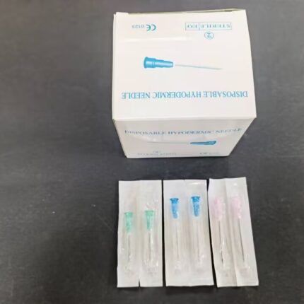
Hypodermic Needle
Disposable hypodermic needles have several essential characteristics for their medical use:
Sterility and Single Use:
They are sterilized (e.g., by EO gas, as shown in the image “STERILE EO”) and designed for single use only to prevent contamination and the transmission of infections.
Material of Construction:
Made of high-quality stainless steel, they are non-toxic and non-pyrogenic.
Tip Design:
They feature a sharp, beveled tip for smooth penetration and maximum patient comfort.
Easy Identification:
The needle hub is color-coded by size (gauge) for quick and easy identification, and can be semi-transparent to allow for visualization of blood flow (flashback).
Compatibility:
They are designed to securely connect to syringes, often via a Luer Lock or Luer Slip connector.
Quality Certifications:
They bear conformity marks such as the CE mark (e.g., “CE 0123” in the image), attesting to their compliance with European safety and quality standards.
Packaging:
Each needle is generally individually sterile packaged to maintain its integrity until use.
