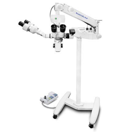
Operation Microscope
Optics & Vision
High Magnification: Magnification ranges from approximately 4x to 40x, providing surgeons with the ability to see very fine details.
Binocular Stereoscopic Vision: Allows for a three-dimensional view of the surgical field, which is crucial for depth perception and performing precise maneuvers.
High-Quality Optics: Features multi-coated optical lenses to enhance light transmission and anti-reflective properties.
Apochromatic Technology: Uses apochromatic correction to ensure that different wavelengths of light focus at the same point, resulting in a clearer and sharper image
High-Power Coaxial Illumination: Provides intense, adjustable light focused along the optical axis, illuminating the surgical field without obstruction.
Adjustable Light Intensity: Allows the surgeon to control the brightness of the illumination to suit the specific procedure and tissue conditions.
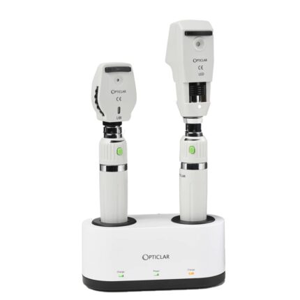
Ophtalmoscope + Rechargeable Retinoscope
- Improved light path and excellent optical performance. ·Imported halogen bulb provides high and adjustable brightness and good color …
- ophthalmoscope; Streak retinoscope; 1Adapt lithium Ion rechargeable handles and USB cable;
- With Rechargeable Battery Handle.
- Smart, portable
- Superior miniaturised optics facilitates entry into even very small diameter pupils; Polarising filter practically eliminates ..
- original optical systems for minimal corneal reflection · Polarized Filter Wide Range of Corrective Lenses · Direct Reading Diopter Indicator.
The portable product has excellent optical performance by improved optical design. Features: With improved optical design and excellent optical performance.
ophthalmoscope head with various diagnostic apertures and filters, and a retinoscope head with a rotatable and variable-length streak for measuring refractive error and astigmatism. Key characteristics include excellent optics, continuously adjustable brightness, and quick, convenient, and safe recharging to protect the battery.
Ophthalmoscope Characteristics
Light Source & Optics: Provides improved light paths and excellent optical performance with imported halogen or LED bulbs for high, adjustable brightness.
Filters: Includes red-free filters to enhance the visibility of retinal details and a polarizing filter for improved contrast.
Lenses: Features a wide range of corrective lenses (e.g., +9D to -10D, with auxiliary options for ±20D) to aid in viewing the retina.
Beams: Offers multiple diagnostic beams, such as a wide-angle beam for general views, a slit beam to assess retinal elevations, and a fixation cross.
Portability: A smart, portable design with a brow rest for user comfort, designed for convenient and mobile examinations.
Retinoscope Characteristics
Illumination: Utilizes a focused light source, often a streak, which can be adjusted to be parallel, convergent, or divergent.
Streak Control: The streak of light rotates 360 degrees and can be moved upward and downward, allowing for precise measurement of the astigmatism axis.
Brightness: Provides continuously adjustable brightness to suit different examination conditions.

Ophthalmic Operating Table
The features of an ophthalmic operating table, such as the one shown, generally include the following:
Specialized Design for Ophthalmology: The table is specifically designed for ophthalmic surgeries and treatments, providing optimal positioning for delicate ocular procedures.
Triple-Section Tabletop: The tabletop is often divided into three sections, with a detachable head section and an integrated drainage system to facilitate eye surgeries.
Durable and Easy-to-Clean Materials: The base and upholstery are made of stainless steel, ensuring good stability, corrosion resistance, and easy cleaning, thus reducing the risk of infection.
Ergonomic Adjustments: The table allows for various position adjustments (height, Trendelenburg, lateral tilt) for patient comfort and surgeon ergonomics, often controlled by a manual unit.
Special Accessories: It is equipped with an ophthalmic headrest for stable positioning of the patient’s head and can include padded arm supports for the surgeon.
Operation: Available in hydraulic or electric options, providing smooth and precise tabletop movement for precise patient positioning.
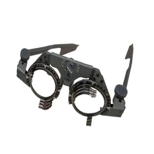
Ophthalmic Trial Frame
The characteristics of an ophthalmic trial frame like the one shown generally include the following:
Material and Design:
Often made of titanium alloy and plastic for lightweight performance.
Simple design for easy fitting.
Lens Capacity:
Holds up to 4 pairs of trial lenses (3 front and 1 back).
Adjustments for a Precise and Comfortable Fit:
Pupillary Distance (PD): Adjust the PD (e.g., 54-70mm) with a single knob.
Lens Axis: Adjust the astigmatic axis by turning a dedicated knob after inserting the lenses.
Temple Angle: Temple angle adjustment knob for optimal positioning (e.g., a tilt angle of approximately 15°).
Temple Length: Adjustable by gently pulling the adjustment part.
Nose Pads: Adjustable in length and angle to fit the patient’s nasal bridge.
Ear Pads: Often equipped with soft ear pads for patient comfort.
Scales and Graduations:
Axis Scale: The axial scale increases counterclockwise along the frame axis, with a marking interval of 5°.
Lenses rotate 360° around the optical axis within the lens frame.
Use:
Suitable for eye examinations in optical stores, hospital ophthalmology departments, schools, and businesses.

Ophthalmic Ultrasound Pachymeter-Biometer
MODEL: SW-1000AP
The device shown is an ophthalmic ultrasound pachymeter-biometer, such as the SW-1000AP or a similar model. Its main features include:
Display: Large color touchscreen with backlight and brightness control, facilitating operation.
Portability: Portable design and lightweight (approximately 1.2 kg for some models), making it easy to carry.
Integrated Features: Integrates a Type A (10 MHz) ultrasound biometer and a Type P (20 MHz) ultrasound pachymeter into a single combined unit.
Printer: Built-in high-speed thermal printer.
Pachymetry: Reliable measurement of central and peripheral corneal thickness with automatic compensation, including detailed pachymetry maps.
Biometrics: Accurate measurements with molecular identification, implant power calculations using common formulas, and automatic or manual measurement modes.
Probe: Uses an LED light probe for biometrics.

Ophthalmic YAG Laser System
MODEL: MD-920
The MD-920 ophthalmic YAG laser system, as shown in the image, has the following specifications:
Laser Specifications:
Laser Type: Nd:YAG, Q-switched
Wavelength: 1064 nm
Mode: Q-switched (multimode)
Pulse Output: Single pulse, double pulse, and triple pulse burst
Pulse Duration: 4.5 ns
Repetition Rate: 2.5 Hz
Cone Angle: 18°
Spot Size: 30 µm
Aiming Beam Wavelength: 633 nm
Aiming Beam Output Power: <0.4 mW
Slit Lamp (Galilean Stereoscopic Microscope) Features:
Provides high clarity and sharpness, avoiding double images.
Five-level drum magnification.
Optional accessories: teaching tube, CCD camera, and tonometer.
Other Features:
Easy setup for installation and maintenance.
Mounted on a mobile rolling stand for transport.
Uses microprocessor control technology with a digital display for easy operation.
Built-in laser protection system to ensure surgical safety.

Ophthalmoscope
An ophthalmoscope is a medical instrument used for examining the eye. Its general characteristics include:
Type:
This is a direct optical ophthalmoscope, portable and battery-powered (2 AA batteries).
Optics and Illumination:
It features a light source, an LED bulb, offering high and adjustable brightness, with good color rendering.
The optical system is designed to minimize glare and ensure clear visibility of the back of the eye, even with small pupils.
It can include different diaphragms and filters (e.g., a green filter that eliminates red) for various observations.
Optical Correction:
A lens correction range (from -20 D to +20 D) is integrated to compensate for ametropia in the examiner or patient.
Design and Ergonomics:
The handle is designed for a comfortable grip and is compatible with various instrument heads.

Optical Biometer
The AL-VIEW optical biometer is an ophthalmic instrument designed for precise eye measurements, particularly for calculating intraocular lens (IOL) fit prior to cataract surgery.
Its main features include:
High-precision measurement: It is capable of measuring and calculating the visual axis and IOL with high accuracy.
Measurement functions: It can measure axial length, keratometry (corneal radii of curvature), and pupillometry.
Automatic operation: The device allows automatic operation without manual manipulation; results are obtained with a simple touch.
Compact design: It incorporates a computer into its design, making it compact and self-contained.
Micron accuracy: It ensures accurate measurement of the eye’s axial length to the micron level.
Biological progression analysis: Biological progression analyses are available.
Built-in thermal printer: It features a built-in thermal printer for printing results.
Connectivity: It can be equipped with USB ports and a network connection to communicate with patient management software.

Optical Coherence Tomograph
The device shown is a Nidek RS-3000 Advance 2 optical coherence tomograph (OCT). Its key features include:
Comprehensive Retinal and Glaucoma Analysis: It offers a complete solution for analyzing retinal pathologies and glaucoma.
SLO-Based Eye Tracker: A scanning laser ophthalmoscope (SLO)-based eye tracker system enables precise image capture with eye movement compensation.
Selectable OCT Sensitivity: Enables B-scan image acquisition even through opacities in the ocular media.
HD Plus Tracing: This feature ensures accurate averaging of up to 120 images for high-resolution, high-contrast images.
OCT Angiography (OCT-A): High-resolution OCT angiographic imaging capability for observing microvascular details of the retina.
Glaucoma Analysis with Normative Database: Includes glaucoma analysis with an extensive normative database (9 x 9 mm).
High Scanning Speed: The device can reach 85,000 A-scans per second, enabling rapid image acquisition.
Automatic Panoramic Imaging: Automatic panoramic imaging capability for OCT angiography up to 12 x 12 mm.
Vibrant Color LCD Display: Equipped with an 8.4-inch color LCD display for real-time data viewing and review.
Comfortable Working Distance: A 35.5 mm working distance ensures comfortable patient positioning.

Optical Lens Cutting Machine
MODEL: SJG-1000
The features of the SJG-1000 (or SJG-1000AK) optical lens cutting machine are as follows:
Tracing and Scanning System:
Optical scanning and centering are positioned as a single system.
Obtain the frame shape in 0.5 seconds.
Real-time imaging guidance to confirm the optimal center position.
Automatic elimination of lens, prism, and artificial parallax effects.
Display and Interface:
7-inch high-resolution (800*600) color touch screen.
Simple operation and low failure rate, requiring little maintenance.
Lens Processing Capabilities:
Suitable for various lens materials, including glass, CR, PC, high-index lenses, and CR-39.
Equipped with various wheels (diamond for glass, V-groove for CR/PC, bevel polishing).
Processes lenses from 22 mm to 80 mm.
Automatic and manual beveling are available.
Grooving width, depth, and position are manually adjustable.
V-shaped and flat polishing are available.
Other Features:
Can be connected simultaneously to two SJG-1000AK automatic milling machines to form a processing center.
3D plotter for accurate lens imaging in 1 second on an 8-inch monitor (for the AK version).
Built-in, easy-to-use blocker (for the AK version).
Four-level adjustable scanning light source (for the AK version).
Compact design for easy placement in a workshop or laboratory.

Optical Lens Trial Case
The optical lens trial case has the following characteristics:
Type of Equipment:
This is a trial lens set (or “Trial Lens Set”), used in optometry and ophthalmology.
Contents:
It contains a full range of spherical lenses (positive and negative), cylindrical lenses (positive and negative), and prisms, as well as accessories such as a trial frame and an occluder.
Use:
This set is essential for determining the necessary visual correction during an eye exam, allowing you to test different lens powers to refine your optical prescription.
Case Materials:
The case is rigid, made of aluminum or a similar material, with a padded interior to protect the lenses.
Organization:
Lenses are generally systematically organized by dioptric power and type (spherical, cylindrical) to facilitate quick selection by the practitioner.

Optical Prisms
The characteristics of optical prisms, such as those shown in the image, are as follows:
Geometric Shape: A prism is a transparent body bounded by two non-parallel plane faces, called refracting faces, and a base. The prisms in the image are individual prisms, often used in ophthalmology.
Light Deflection: The main characteristic of a prism is its ability to deflect light. When a light ray passes through a prism, it is deflected toward the base of the prism.
Chromatic Dispersion: Due to the variation in the refractive index of the prism material depending on the wavelength of light, a prism can disperse white light into its spectral colors (dispersion phenomenon).
Total Internal Reflection: In certain configurations, prisms can use total internal reflection to deflect light with minimal loss, often more effectively than traditional mirrors.
Use in Ophthalmology: Prisms are commonly used in ophthalmology to correct binocular vision problems such as diplopia (double vision) or symptomatic heterophoria by shifting the image to match the eye’s position.
Prismatic Power: The power of a prism is measured in prism diopters (Δ) and indicates the prism’s ability to deflect light. The higher the diopter value, the greater the deviation.
