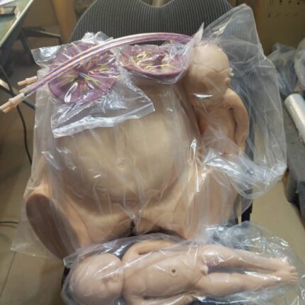
Birthing Pelvis with 2 Babies (Male and Female)
The birthing pelvis is the female bony structure essential for childbirth, providing both structural support and a dynamic passageway for fetal delivery.
Anatomical Composition
-
Comprises four fused bones:
-
Paired iliac bones (forming the pelvic girdle)
-
Sacrum (posterior base of the spine)
-
Coccyx (tailbone)
-
-
Connects the spine to the lower limbs, ensuring stability and weight distribution.
Shape & Structural Divisions
-
Funnel/hourglass shape with distinct regions:
-
Upper flare (false pelvis): Wider, supports abdominal organs.
-
Lower flare (true pelvis): Narrower, forms the birth canal.
-
-
Superior strait: The narrowing between these regions, a critical landmark for obstetric assessment.
Obstetric Straits & Key Measurements
The pelvis features three key straits, each with critical diameters for fetal passage:
-
Upper strait:
-
Promontoretropubic diameter (anteroposterior measurement).
-
-
Middle strait:
-
Median transverse diameter (widest point between ischial spines).
-
-
Lower strait:
-
Interspinous diameter (between ischial tuberosities).
-
-
These measurements determine cephalopelvic disproportion (CPD) and delivery feasibility.
Dynamic Adaptability
-
Not rigid: Hormonal changes (relaxin, progesterone) increase ligament laxity late in pregnancy.
-
Pubis symphysis and sacroiliac joints slightly widen, expanding the birth canal by up to 10–15%.
Role in Childbirth
-
Guides fetal descent: The baby navigates through the pelvic inlet, mid-cavity, and outlet.
-
Facilitates rotation: Bone contours help the fetus rotate into optimal positioning (e.g., occiput anterior).
-
Enables vaginal delivery: Optimal pelvic dimensions and adaptability prevent obstructed labor.

Birthing Pelvis with 2 Babies (Male and Female)
The Birthing Pelvis: Structure and Function in Childbirth
The female birthing pelvis serves as the crucial bony framework that enables successful vaginal delivery. This specialized structure combines anatomical precision with dynamic adaptability to facilitate the birth process.
Bony Architecture:
-
Composed of four interconnected bones:
-
Paired ilium bones (forming the sides)
-
Sacrum (posterior wall)
-
Coccyx (tailbone)
-
-
Forms a complete pelvic ring connecting the spine to the femurs
-
Provides structural support while allowing necessary movement
Morphological Features:
-
Distinctive hourglass configuration with:
-
Expanded false pelvis (greater pelvis) superiorly
-
Constricted true pelvis (lesser pelvis) inferiorly
-
Demarcated by the pelvic brim (superior strait)
-
-
Exhibits characteristic female adaptations:
-
Wider subpubic angle (>90°)
-
Broader, more circular inlet
-
Shorter sacral curvature
-
Obstetric Dimensions:
Three critical planes determine fetal passage:
-
Pelvic Inlet (Superior Strait):
-
Key diameter: Promontoretropubic (11.5-13 cm)
-
-
Midpelvis:
-
Crucial measurement: Median transverse (10.5 cm)
-
-
Pelvic Outlet:
-
Important span: Interspinous (10-11 cm)
-
Dynamic Adaptations:
-
Hormonally-mediated changes (relaxin, progesterone):
-
Increased sacroiliac joint mobility
-
Pubic symphysis widening (2-3 mm)
-
Ligamentous relaxation
-
-
Permits up to 10-15% expansion of birth canal dimensions
Parturition Mechanics:
-
Fetal Engagement: Head enters pelvic inlet
-
Descent: Progressive movement through planes
-
Rotation: Internal adjustment to navigate contours
-
Expulsion: Final passage through outlet

Blocks for half pins.
Connection to Frame: These blocks connect the half pins, which are inserted into the bone, to a central ring or frame component of the external fixator.
Centers: They typically attach to a ring using a post and a one-hole cube, with a hexagonal set screw used to lock the centering sleeve in place.
Half Pin Attachment: They facilitate the attachment of half pins, allowing for the creation of a more rigid construct.
Size and Hole Configuration: Blocks are available with different numbers of holes (e.g., 2, 3, 4 holes) to accommodate various fixation needs.
Centering Sleeves: Centering sleeves are used to ensure that half pins are properly aligned within the block, providing a more stable connection to the half pin itself.
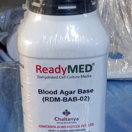
Blood Agar Base
ReadyMED™ Blood Agar Base (RDM-BAB-02)
Manufactured by Chaitanya Agro Biotech Pvt. Ltd.
Product Overview
A high-quality dehydrated blood agar base medium designed for the cultivation of fastidious microorganisms and hemolysis observation. Ideal for clinical, pharmaceutical, and research laboratories.
Key Features
-
Universal base medium for preparing blood agar plates
-
Optimal nutrition for fastidious organisms (streptococci, pneumococci, etc.)
-
Clear hemolysis visualization when supplemented with 5-10% blood
-
Ready-to-prepare dehydrated formulation
Technical Specifications
| Parameter | Specification |
|---|---|
| Product Code | RDM-BAB-02 |
| Form | Free-flowing powder |
| Typical Composition | Peptones, beef extract, NaCl, agar |
| pH (prepared) | 7.3 ± 0.2 |
| Sterilization | Requires autoclaving (121°C for 15 mins) |
| Recommended Supplement | 5-10% defibrinated blood |
Applications
-
Isolation of pathogenic streptococci
-
Hemolysis pattern studies (α, β, γ)
-
Routine culture of fastidious organisms
-
Clinical microbiology diagnostics
Manufacturer Details
Chaitanya Agro Biotech Pvt. Ltd.
-
Registered Corporate Identity Number (CIN) provided on label
-
Customer support contacts included
-
Website for product information
Preparation Protocol
-
Suspend 40g in 1L distilled water
-
Heat to boiling with stirring
-
Autoclave at 121°C for 15 minutes
-
Cool to 45-50°C; add sterile blood aseptically
-
Pour plates under sterile conditions
Quality Assurance
-
Manufactured under strict quality controls
-
Batch-to-batch consistency
-
Complies with international microbiological standards

Blood Agar Base Culture Medium
ReadyMED™ Blood Agar Base (RDM-BAB-02) – Product Specifications
Product Overview
A dehydrated culture medium formulated for preparing blood agar plates and cultivating fastidious microorganisms. Manufactured by Chaitanya Agro Bio-Tech Pvt. Ltd.
Key Features & Applications
Functional Properties
✔ Nutrient-Rich Formula – Supports growth of demanding pathogens
✔ Versatile Usage – Can be used:
-
As a base for blood agar (5-10% defibrinated blood addition)
-
As a general-purpose medium (without blood)
Primary Applications
-
Isolation & Cultivation of Fastidious Bacteria:
-
Streptococcus spp.
-
Neisseria spp.
-
-
Hemolysis Studies:
-
Differentiation of Listeria species
-
CAMP test for Streptococcus agalactiae identification
-
-
Clinical & Diagnostic Use:
-
Blood cultures
-
Respiratory/sputum specimens
-
Composition Highlights
-
Peptones & Yeast Extract – Provide essential nitrogen/vitamin sources
-
Sodium Chloride – Maintains osmotic balance
-
Agar – Solidifying agent (1.5-2.0% typical concentration)
Technical Specifications
-
Form: Free-flowing powder
-
pH (after reconstitution): 7.3 ± 0.2
-
Sterilization: Autoclave at 121°C for 15 mins
-
Storage: Keep sealed at 10-30°C; avoid humidity
Preparation Guide
-
Suspend 40g in 1L distilled water
-
Heat to dissolve completely
-
Sterilize & cool to 45-50°C
-
Add 5-10% sterile blood (if required)
-
Pour into plates
Quality Assurance
-
Batch-Tested for microbial growth performance
-
ISO Certified manufacturing
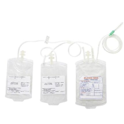

Blood Bag Shaker
The Smart Weighing Instrument for Blood Collection offers several key features:
Weighing and Control Functions:
It is equipped with load weighing, bag weight detection, volume control, pan vibration, and automatic restart.
Gentle Shaking:
A gentle shaking mode is integrated to improve the survival of effective components in the blood.
Automatic Shaking Stop and Restart:
Stirring stops automatically when the bags are removed during collection and restarts once the bags are replaced.
Accuracy and Display:
It offers high measurement accuracy thanks to a taring and calibration function, and features a clear LED display for blood volume measurement.
Alarms and automatic shutoff:
The device automatically sounds an alarm when the preset volume is reached and stops stirring when the collection volume is close to the preset value (e.g., within 10 ml of the target value) for accurate measurement.
Design and durability:
It is designed for ease of use and robustness, with a corrosion-resistant metal body for increased durability in medical environments.

Blood Bank Refrigerator
The blood bank refrigerator
This equipment is specifically designed for the secure storage of blood and its components, as well as laboratory reagents and biological products, at controlled temperatures (generally between +2°C and +6°C). It is equipped with multiple safety features to ensure optimal preservation of blood products.
Volume: Available in 200L and 400L

Blood collection chair
Blood collection chair, used for blood collection, transfusions, or blood donations.
Materials:
It is usually made of mild steel with a PVC coating on the seat and backrest, providing durability and ease of cleaning.
Design:
It is designed for patient comfort, with a padded backrest and seat, and armrests.
Specific Features:
It is equipped with an adjustable IV pole (infusion pole), visible on the side.
Stability and Durability:
The frame is often welded and powder-coated for long life and safe use in a hospital environment.
Adjustability:
Some models may offer backrest and knee adjustments to suit the patient’s needs.

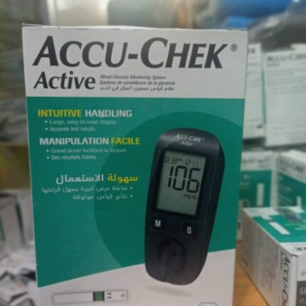
Blood Glucose Meter
The OneTouch Ultra 2 blood glucose meter has several notable features:
Accuracy and Speed of Results:
It provides accurate results in 5 seconds.
Storage Capacity:
The meter can store the most recent 500 blood glucose and control solution test results, displayed in chronological order.
Display Features:
It features a large, easy-to-read display with a backlight for nighttime use.
Meal Tagging:
The system allows you to tag test results before and after meals, providing separate averages for these periods and helping you understand the impact of diet on blood glucose levels.
Ease of Use:
It is designed with two-way scrolling and easy-to-use buttons.
Connectivity:
It is equipped with a download port for data retrieval.
Sampling options:
It allows blood to be drawn from various test sites, including the arm where there are fewer nerve endings.

Blood Glucose Test Strips
OneTouch Ultra Blood Glucose Test Strips feature the following features
DoubleSure™ Technology: Automatically checks each blood sample twice to confirm the result, ensuring proven accuracy.
FastDraw™ Capillary Action: Automatically draws blood into the strip for quick and easy sample collection.
Small Sample Size: Requires only a small drop of blood, making testing less painful.
Fast Results: Provides results in as little as five seconds.
Visual Confirmation: Makes it easy to see when there is enough blood for an accurate reading.
Compatibility: Designed for use with the OneTouch Ultra family of meters, including the OneTouch Ultra 2, UltraMini, UltraSmart, UltraLink, and the OneTouch Ping Meter Remote.
Alternative testing options: Allows glucose levels to be tested on sites other than the fingertip (e.g., the forearm), after consulting a healthcare professional.
